
Get a Free Second Opinion

Cardiology Department with 24/7 Support

Cardiology Department with 24/7 Support

Best Cardiology Hospital in Hyderabad
- Cardiologists with 35+ Years of Expertise
- World-Class Facilities for Advanced Cardiac Care
- 24/7 Fully Equipped Cardiac ICUs
- Leader in Minimally Invasive Cardiac Surgeries
- Leading Center for Heart-Lung Transplantations
- Highest Number of Cardiac Procedures with High Success Rates
- Advanced Electrophysiology Services
- Cutting-Edge Diagnostic & Cath Lab Technologies
- Specialized Pediatric Cardiac Interventions
- Dedicated Patient Coordinators and International Support Services
At Yashoda Heart Institute, we are committed to providing unparalleled cardiac care through a comprehensive multidisciplinary approach. Known as one of the best Cardiology Hospitals in Hyderabad and among the top Heart hospitals in India, we offer extensive diagnostic, medical, and surgical cardiology services. Our state-of-the-art heart clinics and highly skilled team of cardiologists, surgeons, and healthcare professionals put us at the forefront of cardiac innovation. We provide advanced treatment and quality care, ensuring that each patient receives the best outcomes and is on the path to a healthy heart.
Center of Excellence
Our Centre of Excellence offers advanced cardiac interventions and specialised treatments for complex coronary artery disease, cardiac rhythm disorders, and structural heart and valvular conditions through minimally invasive interventions, complex coronary procedures, and advanced electrophysiology studies. Supported by Optical Coherence Tomography (OCT) and Intravascular Ultrasound (IVUS), we deliver precise diagnosis, optimised treatment planning, and precision-driven cardiac care.
- TAVI
- TAVR
- Heart Transplant
- Structural valve intervention
- Electrophysiology
- Radiofrequency ablation (RFA)
- Device implantation
- Complex Percutaneous Coronary Intervention (PCI)
- Image-guided Percutaneous Transluminal Coronary Angioplasty (PTCA)
- Optical Coherence Tomography (OCT) and Intravascular Ultrasound (IVUS)
- Rotablation (rotational atherectomy)
- Orbital atherectomy (OA)
- Aortic aneurysm
- Mitral transcatheter edge-to-edge repair (TEER)
- Rescue angioplasty
- Left Ventricular Hypertrophy (LVH)
- Left Ventricular Assist Device (LVAD)
- Mitral valve-in-valve (ViV)
Pioneering Excellence in Cardiac Care
- Successfully performed the first inter-state Heart Transplantation in Telangana and AP.
- Performed the first-ever combined heart-lung transplantation successfully in Telangana and AP.
- Recognized as one of the world’s leading Comprehensive Heart & Lung Transplantation Centers, achieving exceptional outcomes in complex procedures.
- The first Dual Source CT with Heart PBV was introduced in India, enhancing diagnostic accuracy and patient care standards.
- Collaborated with surgeons from Duke University, USA, to perform PTE procedures in Pulmonary Embolism, ensuring world-class treatment standards.
- Offers state-of-the-art facilities for Ventricular Assist Devices (VADs), Pacemakers, and ICD Implantations, ensuring comprehensive cardiac care.
- Pioneers in Awake ECMO procedures and Air Ambulance Transport Facilities, ensuring swift and effective critical care response.
- First in the region to successfully perform WATCHMAN Device Implantation, facilitating stroke prevention in atrial fibrillation patients.
- First in the region to successfully perform Transcatheter Mitral Valve Replacement, offering minimally invasive solutions for heart valve disorders.
- Performs the maximum number of cardiac procedures (20,000 annually, including 600 PTCAs monthly), underscoring leadership in cardiac interventions.
- Recognized for excellence in Complex Cardiothoracic and Minimally Invasive Cardiac Surgeries, offering advanced treatment for various heart and lung conditions.
Why Choose Yashoda Hospitals?
The Cardiology Department at Yashoda Hospitals is equipped with state-of-the-art technologies and equipment, ensuring the highest standard of cardiac care.
- Advanced Diagnostic Tools: We offer cutting-edge imaging technologies such as CT Coronary Angiogram, Cardiac MRI, and Radionuclide Studies. Our facilities also include the latest 2D/3D ECHO, TEE, DSE, Head-Up Tilt Test, HOLTER, and ABPM for comprehensive cardiac evaluation.
- Specialized Cath Labs: Our Hybrid Cath Lab and dedicated Neuro Intervention Cath Lab are equipped with the most advanced tools, including flat-panel imaging technology, IVUS, OCT, NIRS IVUS, and 3D Mapping for precise diagnosis and treatment.
- Innovative Treatment Options: Our department specializes in coronary, structural, and peripheral interventions, as well as electrophysiology diagnosis and intervention. We provide image-guided complex coronary interventions, including OCT/IVUS-guided procedures, Left Main and Bifurcation CTO treatments, Post-bypass Interventional Complex Angioplasty, and support with devices like Impella and PulseCath-assisted ECMO.
- Cutting-Edge Procedures: We offer advanced treatments such as TAVR, TMVR, MITRA CLIP, LAA Appendage Closure, and the MICRA leadless pacemaker. Our electrophysiology team performs advanced procedures using 3D mapping for VT and atrial fibrillation, including renal denervation and advanced ETS & RFA.
- Comprehensive Heart Failure Management: Our expertise extends to heart failure device implantations and structural interventions, including TAVI, TMVR, and MITRA CLIP procedures.
Advanced Diagnostic and Preventive Cardiac Care
Our doctors at Yashoda Heart Institute can diagnose and treat heart and vascular problems using the latest technology and skills that are unmatched skills. Our team of interventional cardiologists specialises in treating and preventing heart disease for any cardiovascular disease suspect seeking medical attention.
Regular Screening Programs and Heart Health Check-ups
Yashoda Hospitals offers regular screening programs and heart health check-ups to ensure early detection and prevention of cardiac conditions. Our preventive and curative cardiology services are designed to maintain and improve heart health for all patients.
By combining advanced technology, specialized expertise, and a commitment to patient-centered care, Yashoda Hospitals stands out as a leader in the field of cardiology.
We offer specialized heart-related health check-up packages, including:
Experts in Cardiac Treatments and Surgeries
With three decades of experience, Yashoda Heart Institute is a leading cardiology hospital in India. We are well-recognised for pioneering procedures like combined heart transplants, advanced infrastructure, and the adoption of cutting-edge technology. Our expertise and commitment to advanced and personalised cardiac care have been demonstrated, resulting in excellent patient outcomes.
Our commitment to pushing the boundaries of medical technology and treatment has allowed us to provide exceptional cardiac care, resulting in our recognition as a top cardiology hospital in India and the achievement of many medical breakthroughs. This hospital offers advanced cardiac care, including access to highly skilled surgeons who perform over 20,000 minimally invasive heart procedures annually.
We use state-of-the-art technology, which allows us to customise cardiac treatments based on individual needs. For patients seeking heart treatment in Hyderabad, Yashoda Heart Institute is the best option, not only because of the expertise of the doctors but also because of the advanced infrastructure available for you. This means you will receive the best possible cardiac care and have the best chance for a better recovery.
Advanced Cardiology Treatments in Hyderabad
- Structural valve interventions
- TAVI
- Transcatheter Aortic Valve Replacement (TAVR)
- TEER
- TMVR (Transcatheter Mitral Valve Replacement)
- Balloon Valvuloplasty
- Transcatheter Pulmonary Valve Replacement (TPVR/TPVI)
- Mitral Valve-in-Valve (ViV)
- Treatment for Paravalvular Leakages
- Aortic Aneurysm Repair
- Endovascular Aneurysm Repair (EVAR)
- Thoracic Endovascular Aortic Repair (TEVAR)
- Hybrid Aortic Arch Repair
- Aortic Root Replacement
- Valve-Sparing Aortic Root Replacement (David Procedure)
- Aortic Dissection Repair (Type A / Type B)
- Treatment for Left Ventricular Hypertrophy (LVH)
- Septal Myectomy
- Alcohol Septal Ablation
- WATCHMAN Device Implant
- Micra Leadless Pacemaker Implantation
- Impella Heart Pump
- Left Main Stenting
- Bifurcation Stenting
- ECMO Support with PulseCath
- Renal Denervation
- Graft Stenting (SVG Stenting)
- Chronic Total Occlusion Percutaneous Coronary Intervention (CTO PCI)
- TriClip / Transcatheter Tricuspid repair
- Rotational Atherectomy (Rotablator)
- FFR-Assisted Procedures
- Pulmonary Thromboendarterectomy (PTE)
- Aortic Root Valve Dysfunction
- Atrial Myxoma Excision
- Atrial Fibrillation Ablation
- Atrial Septal Defect Closure
- CRT-D
- CRT-P
- Carotid Artery Stenting
- CABG
- Electrophysiology Procedures
- Heart Transplant
- Implantable Cardioverter Defibrillator (ICD)
- Mitral Valve Clip
- Mitral Valve Replacement
- Open Heart Surgery
- Patent Ductus Arteriosus (PDA) Closure
- Permanent Pacemaker Implants (PPI)
- PTCA (Percutaneous Transluminal Coronary Angioplasty)
- Radiofrequency Ablation
- Tetralogy of Fallot Repair
- Treatment for Heart Failure
- Treatment for Cardiac Tumours (Myxoma Excision)
- Ventricular Septal Defect (VSD) Closure
Preview: Structural valve interventions are advanced, minimally invasive, catheter-based procedures used to repair or replace diseased heart valves without the need for conventional open-heart surgery. These procedures offer minimal blood loss due to fewer incisions, often resulting in reduced procedural complications, a shorter hospital stay, and faster recovery.
These include:
- TAVI/TAVR
- TEER (MitraClip)
- TMVR
- Balloon Mitral Valvotomy
- Tricuspid Valve Repair/Replacement
- TPVI
- Valve-in-Valve Procedures
- Paravalvular Leak Closure
Benefits:
- Minimally invasive treatment with smaller incisions
- Faster recovery and shorter hospital stay
- Reduced pain and surgical trauma
- Lower risk of complications in eligible patients
- Improved heart valve function and blood flow
- Relief from symptoms such as breathlessness and fatigue
- Quicker return to daily activities
- Suitable for elderly and high-risk patients
- Enhanced quality of life and overall heart health
- Effective alternative to open-heart surgery in selected cases
Preview: TAVI (Transcatheter Aortic Valve Implantation) is a minimally invasive heart procedure used to replace a narrowed or damaged aortic valve instead of performing an open-heart surgery. It is commonly recommended for patients with severe aortic stenosis, especially elderly or high-risk patients.
Surgical Steps:
- Suitable anaesthesia is given to the patient
- A small catheter is inserted through a blood vessel, usually in the groin
- The catheter is guided towards the heart using imaging technology
- The new artificial valve is positioned inside the damaged aortic valve
- Valve function is checked carefully before completing the procedure
- The catheter is carefully removed after valve placement
- The patient is monitored in ICU or recovery area for observation
Benefits:
- Minimally invasive procedure
- No need of an open heart surgery
- Reduced pain and blood loss
- Faster recovery and early mobilization
- Shorter hospital stay
- Lower risk of complications in selected patients
- Suitable for elderly and high surgical-risk patients
Read more about –Transcatheter Aortic Valve Implantation
Preview: Transcatheter Aortic Valve Replacement (TAVR), or Transcatheter Aortic Valve Implantation (TAVI), is a minimally invasive procedure replacing a narrowed aortic valve in high-risk patients by implanting a prosthetic valve via catheter, which expands over the existing valve and functions immediately, typically completed in two hours.
Surgical Steps:
- Prior to TAVR, patients must discuss medication adjustments, fasting requirements, and allergies with their treatment team to ensure proper procedure preparation.
- During TAVR, a team monitors the heart while a catheter inserted through the leg or chest and guided by imaging delivers a prosthetic valve to the aortic valve, which is then expanded into place, sometimes with a balloon.
- After TAVR, patients must monitor for heart failure symptoms, keep the procedure site clean and dry, take blood-thinning medication, and take antibiotics to reduce infection risk.
Benefits:
- Minimally invasive procedure performed in the cath lab
- No general anaesthesia is required
- No big scar after the procedure
- A short hospital stay of 3-4 days
- Quick recovery and return to normal life
- Lower risk for patients with serious health conditions
Read more about – Transcatheter Aortic Valve Replacement (TAVR)
Preview: TEER (Transcatheter Edge-to-Edge Repair) is a minimally invasive heart procedure used to treat leaking heart valves, especially the mitral valve, without open-heart surgery. It helps improve valve function by clipping together a portion of the valve leaflets to reduce leakage (regurgitation). MitraClip is the specific medical device used to perform the TEER procedure on the mitral valve.
Surgical Steps:
- Suitable anaesthesia is given to the patient
- A catheter is inserted through a blood vessel, usually via the groin
- The catheter is guided towards the heart using advanced imaging technology
- A specialized clip device is positioned across the leaking valve
- The valve leaflets are clipped together to reduce leakage
- Valve function is checked carefully before completing the procedure
- The catheter is removed and the patient is shifted for recovery monitoring
Benefits:
- Minimally invasive alternative to open-heart surgery
- Smaller incision with less pain and blood loss
- Faster recovery and shorter hospital stay
- Improved symptoms like breathlessness and fatigue
- Reduced risk in elderly or high surgical-risk patients
- Better heart function and quality of life
Read more about – Transcatheter Edge-to-Edge Repair
Preview: Transcatheter Mitral Valve Replacement (TMVR) is an advanced minimally invasive heart procedure used to replace a damaged mitral valve without performing open-heart surgery. It is usually recommended for patients with severe mitral valve disease who are considered high risk for conventional surgery.
How is TMVR done?
- A thin catheter is inserted through a blood vessel or small chest incision
- The replacement valve is guided to the heart using advanced imaging
- The new valve is positioned inside the damaged mitral valve
- Once placed correctly, the new valve starts functioning immediately
- The procedure is minimally invasive and usually offers faster recovery compared to open surgery
Benefits:
- Improves blood flow through the heart
- Reduces backflow/leakage from the mitral valve
- Helps the heart pump blood more efficiently
- Reduces strain on the heart chambers
- Improves oxygen supply to the body
- Helps relieve symptoms of heart failure
- Supports better overall cardiac performance and endurance
Preview: This surgery uses a catheter-mounted balloon to stretch the heart valves wide open in patients with age-related nerve stiffness, inflammation, and congenital defects.
Types of Balloon Valvuloplasty:
Balloon valvuloplasty is a minimally invasive catheter-based procedure that opens the narrowed heart valves through several categories, such as:
- Tricuspid valvotomy
- Pulmonary valvotomy
- Aortic valvotomy
- Percutaneous mitral valvotomy
Post-Surgical Care:
- Avoid heavy lifting for a week
- Engage in light walking
- Maintain a healthy, well-balanced diet
- Monitor for complications like insertion site issues or chest pain
- Regular cardiologist follow-ups
Preview: Transcatheter Pulmonary Valve Replacement (TPVR) is a minimally invasive procedure used to replace a damaged or dysfunctional pulmonary valve without open-heart surgery. It is commonly performed in patients with congenital heart diseases who have previously undergone surgical repair and later develop pulmonary valve stenosis or regurgitation.
Surgical Steps:
- A catheter is inserted through a blood vessel, usually in the groin.
- The replacement valve is mounted on a stent and is guided to the pulmonary valve’s position.
- The new valve is expanded within the diseased valve or conduit.
- The valve begins functioning immediately to restore normal blood flow.
Benefits:
- Avoids repeat open-heart surgery
- Minimally invasive approach
- Shorter hospital stay
- Faster recovery
- Improved valve function and blood flow
- Relief from symptoms such as fatigue and breathlessness
Preview: Mitral Valve-in-Valve (ViV) is a minimally invasive transcatheter procedure used to repair a failed or deteriorated surgical bioprosthetic mitral valve without the need for repeat open-heart surgery. It is often recommended for patients who are at high surgical risk or have previously undergone mitral valve replacement.
Surgical Steps:
- A catheter is inserted through a blood vessel or a small incision.
- The replacement valve is guided to the heart and positioned inside the failing bioprosthetic mitral valve.
- The new valve is expanded, anchoring securely within the existing valve frame.
- The new valve immediately takes over the function of regulating blood flow through the mitral valve.
Benefits:
- Avoids repeat open-heart surgery
- Minimally invasive approach
- Shorter hospital stay
- Faster recovery and return to normal activities
- Reduced postoperative complications in high-risk group
- Symptomatic relief and improved valve functionality
- Most common treatment for failed bioprosthetic mitral valves
Read more about –Mitral Valve-in-Valve (ViV)
Preview: Surgical treatments for paravalvular leak are designed to seal abnormal gaps around prosthetic heart valves, restoring efficient blood flow and relieving symptoms such as breathlessness, fatigue, and heart failure. Depending on the severity and complexity of the leak, treatment may involve catheter-based closure, surgical repair, or valve replacement procedures.
Treatments include:
- Surgical Paravalvular Leak Repair
- Repeat Valve Replacement
- Valve-in-Valve Procedures
Preview: The treatment for aortic aneurysms aims to prevent rupture by strengthening or replacing the weakened section of the aorta. Depending on the aneurysm’s location and severity, treatment may involve minimally invasive endovascular techniques or open surgical repair to ensure long-term vascular stability and patient safety.
These include:
- Endovascular Aneurysm Repair (EVAR)
- Thoracic Endovascular Aortic Repair (TEVAR)
- Open Aortic Aneurysm Repair
- Hybrid Aortic Repair
- Aortic Root Replacement (for aneurysms involving the aortic root)
- Valve-Sparing Aortic Root Replacement (David Procedure) in selected patients
Read more about –Aortic Aneurysm Repair
Preview: Endovascular Aneurysm Repair (EVAR) is a minimally invasive vascular procedure used to treat abdominal aortic aneurysms by reinforcing the weakened portion of the aorta with a stent graft.
Surgical Steps:
- A catheter is inserted through blood vessels in the groin
- A stent graft is guided to the aneurysm in the abdominal aorta
- The graft is expanded to seal and reinforce the weakened area
- Blood flow is redirected safely through the graft
- The catheter is removed after confirming proper graft placement
Benefits:
- Helps prevent rupture of the abdominal aorta
- Restores safe blood flow through the aorta
- Minimally invasive alternative to open surgery
- Reduced blood loss and surgical trauma
- Faster recovery and shorter hospital stay
- Useful in selected patients with abdominal aortic aneurysm
Preview: TEVAR is a minimally invasive procedure used to treat diseases affecting the thoracic aorta, including aneurysms, dissections, and traumatic injuries.
Surgical Steps:
- A catheter is inserted through a blood vessel in the groin
- A stent graft is guided to the diseased portion of the aorta
- The graft is expanded to reinforce and seal the damaged area
- Blood flow is redirected safely through the repaired segment
Benefits:
- Minimally invasive alternative to open surgery
- Reduces risk of aortic rupture
- Improves blood flow through the aorta
- Shorter hospital stay and faster recovery
- Reduced surgical trauma and blood loss
Preview: Hybrid arch repair is an advanced procedure combining open surgery and endovascular techniques to treat complex diseases involving the aortic arch.
Surgical Steps:
- Blood vessels supplying the brain may be surgically rerouted
- A stent graft is placed in the diseased aortic arch
- Open surgical and catheter-based techniques are combined for safer repair
- Blood flow through the aorta and branch vessels is restored
Benefits:
- Enables treatment of complex aortic arch disease
- Reduces risk of aortic rupture and complications
- Maintains blood flow to the brain and vital organs
- Less invasive than extensive open aortic surgery in selected patients
- Supports improved long-term cardiovascular health
Preview: Aortic root replacement is an advanced surgical procedure used to replace a diseased or enlarged aortic root, helping prevent serious complications such as aortic rupture while preserving or restoring normal heart and aortic function.
Surgical Steps:
- The surgery is performed under general anaesthesia
- The whole procedure is performed assisted by a heart-lung machine.
- The diseased or enlarged aortic root is removed.
- A synthetic graft is used to replace the affected portion of the aorta.
- Depending on the condition of the aortic valve, it may either be replaced along with the root or preserved (valve-sparing root replacement).
- The coronary arteries are reattached to the new graft to restore normal blood flow to the heart.
Benefits:
- Prevents life-threatening aortic rupture or dissection
- Restores the normal structure and function of the aortic root
- Improves blood flow from the heart
- Treats associated aortic valve disease if present
- Reduces the risk of future aortic complications
Preview: Valve-Sparing Aortic Root Replacement (David Procedure) is an advanced heart surgery performed to treat enlargement or disease of the aortic root while preserving the patient’s natural aortic valve. The procedure helps maintain normal heart valve function and avoids the need for an artificial valve in selected patients.
Surgical Steps:
- The surgery is performed under general anaesthesia.
- The diseased or enlarged portion of the aortic root is removed
- The patient’s natural aortic valve is preserved carefully
- A synthetic graft is used to reconstruct the aortic root
- The preserved valve is reimplanted within the new graft to restore normal blood flow
Benefits:
- Preserves the patient’s natural aortic valve
- Maintains more natural heart valve function
- Improves blood flow from the heart to the body
- Reduces the need for lifelong blood-thinning medications in many patients
- Helps prevent progression of aortic root disease and complications
- Provides durable long-term cardiac function
- Improves quality of life and overall cardiovascular health
Preview: Aortic dissection repair is an emergency cardiovascular procedure performed to treat a tear in the inner wall of the aorta, the body’s main blood vessel. The condition can become life-threatening if not treated promptly.
Surgical Steps:
- The damaged portion of the aorta is identified using advanced imaging
- Type A dissections usually require open-heart surgery
- Type B dissections may be treated with minimally invasive endovascular techniques
- The torn section of the aorta is repaired or replaced with a synthetic graft
- Blood flow through the aorta is restored safely
Benefits:
- Prevents rupture of the aorta
- Restores stable blood flow from the heart to the body
- Reduces strain on the heart and major blood vessels
- Helps protect vital organs from reduced blood supply
- Improves long-term cardiovascular stability and survival
Preview: Treatment for left ventricular hypertrophy (LVH) focuses on addressing the underlying cause of the heart muscle thickening and preventing further progression of the condition. Depending on the severity of LVH and its cause, treatment may range from medications and lifestyle modifications to advanced interventional or surgical procedures.
Surgical interventions include:
- Septal Myectomy
- Alcohol Septal Ablation
- Aortic Valve Replacement (SAVR or TAVI/TAVR)
- ICD Implantation
Preview: Septal myectomy is an advanced cardiac surgery performed to treat hypertrophic obstructive cardiomyopathy (HOCM), a condition in which the heart muscle becomes abnormally thick and obstructs blood flow from the heart.
Surgical Steps:
- The procedure is performed under general anaesthesia.
- The chest is opened to access the heart
- The surgeon removes a portion of the thickened heart muscle (septum)
- The obstruction to blood flow is relieved
- Normal blood flow from the heart improves after surgery
Benefits:
- Improves blood flow out of the heart
- Reduces obstruction caused by thickened heart muscle
- Enhances heart-pumping efficiency
- Relieves symptoms such as chest pain, breathlessness, and dizziness
- Reduces strain on the heart
- Improves exercise capacity and daily functioning
- Helps improve long-term cardiac performance and quality of life
Preview: Alcohol septal ablation is a minimally invasive cardiac procedure used to treat hypertrophic obstructive cardiomyopathy (HOCM), where the heart muscle becomes abnormally thick and blocks blood flow from the heart.
Surgical Steps:
- A thin catheter is inserted through a blood vessel
- The catheter is guided to the artery supplying the thickened heart muscle
- A controlled amount of alcohol is injected into the targeted area
- The excess thickened muscle gradually shrinks
- Blood flow from the heart improves as the obstruction reduces
Benefits:
- Improves blood flow out of the heart
- Reduces obstruction caused by thickened heart muscle
- Enhances overall efficiency of the heart
- Helps relieve chest pain, breathlessness, dizziness, and fainting episodes
- Reduces strain on the heart
- Minimally invasive with shorter recovery time
- Improves exercise tolerance and quality of life in suitable patients
Preview: WATCHMAN Device Implant / LAA Occlusion is a minimally invasive cardiac procedure used to reduce the risk of stroke in patients with atrial fibrillation (AFib). The procedure helps prevent blood clot formation in the left atrial appendage (LAA), a small pouch in the heart where clots commonly develop in AFib patients. (patients with irregular & rapid rhythm)
Surgical Steps:
- A thin catheter is inserted through a vein in the groin
- The catheter is guided to the heart using advanced imaging
- The WATCHMAN device is positioned inside the left atrial appendage
- The device seals the appendage to prevent clot formation
- Over time, heart tissue grows over the device naturally
Benefits:
- Reduces the risk of stroke in AFib patients
- Minimally invasive alternative to open-heart surgery
- Helps reduce long-term dependence on blood thinners
- Lower risk of bleeding complications from anticoagulant medications
- Shorter hospital stay and faster recovery
- Improves long-term heart and vascular safety for eligible patients
Read more about – WATCHMAN Device Implant
Preview: Micra leadless pacemaker implantation is an advanced minimally invasive procedure used to treat slow or irregular heart rhythms. Unlike traditional pacemakers, the Micra device is very small and does not require wires (leads) or a surgical chest incision.
Surgical Steps:
- A thin catheter is inserted through a vein in the groin
- The Micra pacemaker is guided into the heart
- The device is attached directly to the heart wall
- Electrical impulses help maintain a regular heartbeat
- The catheter is removed after successful placement
Benefits:
- No chest incision or visible scar
- No pacemaker wires or leads
- Lower risk of infection and lead-related complications
- Minimally invasive with faster recovery
- Improved comfort and mobility for patients
- Provides reliable and effective heart rhythm control
- Suitable for selected patients requiring pacemaker support
Preview: Impella is a minimally invasive heart support device used in critically ill patients whose heart is unable to pump blood effectively. It temporarily helps maintain blood circulation during severe heart failure, high-risk cardiac procedures, or cardiogenic shock.
Surgical Steps:
- A thin catheter-mounted pump is inserted through a blood vessel
- The device is guided into the heart under imaging guidance
- The pump helps circulate blood from the heart to the body
- It temporarily supports heart function while treatment is continued
Benefits:
- Improves blood circulation throughout the body
- Reduces workload on the weakened heart
- Supports vital organs with better oxygen supply
- Helps stabilize critically ill cardiac patients
- Useful during high-risk cardiac interventions
Preview: A left main stent (percutaneous coronary intervention) is a minimally invasive procedure to open blockages in the left main coronary artery (the critical vessel supplying blood to most of the heart). It is a vital, high-stakes alternative to open-heart bypass surgery (CABG) for carefully selected patients.
Surgical Steps:
- A thin catheter is inserted through a blood vessel in groin
- The catheter is guided to the blocked left main coronary artery
- Balloon angioplasty may be performed to open the blockage
- A coronary stent is placed to restore blood flow
- Blood flow to the heart muscle is assessed before completing the procedure
Benefits:
- Restores blood flow to a major heart artery
- Improves oxygen supply to the heart muscle
- Reduces chest pain and cardiac symptoms
- Helps improve heart pumping efficiency
- Minimally invasive with faster recovery
- Useful in selected high-risk coronary artery disease patients
Preview: Bifurcation stenting is an advanced coronary angioplasty technique used to treat blockages occurring at the division point of a coronary artery into two branches.
Surgical Steps:
- A catheter is inserted through a blood vessel
- Specialized guidewires are passed into both artery branches
- Balloons are used to widen the narrowed segments
- One or more stents are positioned across the bifurcation blockage
- Blood flow in both branches is restored and assessed
Benefits:
- Treats complex coronary artery branch blockages
- Restores smooth blood flow to the heart muscle
- Improves success of complex angioplasty procedures
- Reduces symptoms such as chest pain and breathlessness
- Minimally invasive with shorter recovery time
- Helps improve overall cardiac function
Preview: PulseCath ECMO support is an advanced life-support system used in patients with severe heart or lung failure. It temporarily takes over the function of the heart and lungs, helping maintain oxygen supply and blood circulation during critical conditions.
Surgical Steps:
- Tubes (cannulas) are inserted into major blood vessels
- Blood is circulated through an external ECMO machine
- The machine adds oxygen and removes carbon dioxide
- Oxygen-rich blood is pumped back into the body
- The system supports the heart and lungs while recovery or further treatment continues
Benefits:
- Provides temporary heart and lung support
- Maintains oxygen delivery to vital organs
- Helps stabilize critically ill patients
- Supports recovery after severe cardiac or respiratory failure
- Useful in emergency and intensive care settings
- Can act as a bridge to recovery, surgery, or transplantation
Preview: Renal denervation is a minimally invasive procedure used to help control high blood pressure that remains uncontrolled despite medications and lifestyle changes. The procedure targets overactive nerves around the kidney arteries that contribute to persistent hypertension.
Surgical Steps:
- A thin catheter is inserted through a blood vessel in the groin
- The catheter is guided to the arteries supplying the kidneys
- Controlled energy is delivered to the surrounding nerves
- The overactive nerve signals contributing to high blood pressure are reduced
- The catheter is removed after the procedure
Benefits:
- Helps lower uncontrolled high blood pressure
- Reduces strain on the heart and blood vessels
- May decrease the risk of stroke, heart disease, and kidney damage
- Minimally invasive with no major surgical incision
- Shorter recovery time compared to open procedures
- Can improve long-term blood pressure management in selected patients
Read more about – Renal Denervation
Preview: Graft stenting is a minimally invasive cardiac procedure used to treat narrowing or blockage in bypass grafts previously placed during coronary artery bypass surgery (CABG).
Surgical Steps:
- A catheter is inserted through a blood vessel
- The blocked bypass graft is identified using imaging guidance
- Balloon angioplasty is performed to open the narrowed graft
- A stent is placed to restore blood flow through the graft
- Blood circulation to the heart muscle is reassessed
Benefits:
- Restores blood flow through blocked bypass grafts
- Improves oxygen supply to the heart muscle
- Reduces chest pain and cardiac symptoms
- Avoids repeat major bypass surgery in selected patients
- Minimally invasive with faster recovery
- Helps improve overall cardiac performance
Preview: Chronic Total Occlusion Percutaneous Coronary Intervention (CTO PCI) is an advanced minimally invasive cardiac procedure used to open completely blocked coronary arteries that have been blocked for a long duration. This procedure helps restore blood flow to the heart muscle and improve overall cardiac function.
Surgical Steps:
- A thin catheter is inserted through a blood vessel in the wrist or groin
- Specialized wires and devices are guided to the completely blocked coronary artery
- The blockage is carefully crossed and opened
- Balloon angioplasty is performed to widen the artery
- A stent may be placed to maintain blood flow to the heart
Benefits:
- Restores blood flow to previously blocked heart arteries
- Improves oxygen supply to the heart muscle
- Reduces chest pain and breathlessness
- Enhances heart pumping efficiency and cardiac performance
- Improves exercise tolerance and daily activity levels
- May reduce the risk of future cardiac complications
- Helps improve overall quality of life in selected patients
Preview: Transcatheter Tricuspid Valve Repair (TriClip) is an advanced minimally invasive cardiac procedure used to treat severe tricuspid valve leakage (tricuspid regurgitation). The procedure helps improve valve function and blood flow without the need for open-heart surgery.
Surgical Steps:
- A thin catheter is inserted through a blood vessel, usually from the groin
- The catheter is guided to the tricuspid valve using advanced imaging
- The TriClip device is positioned to bring the valve leaflets closer together
- The valve leakage is reduced, improving blood flow through the heart
- The catheter is removed after successful device placement
Benefits:
- Reduces tricuspid valve leakage
- Improves forward blood flow through the heart
- Decreases strain on the right side of the heart
- Helps relieve symptoms such as swelling, fatigue, and breathlessness
- Improves overall cardiac efficiency and quality of life
- Minimally invasive with faster recovery compared to open-heart surgery
- Suitable for high-risk patients who may not tolerate major surgery well
Preview: Rotational Atherectomy and Orbital Atherectomy are minimally invasive cardiac procedures used to treat severely calcified or hardened blockages in the coronary arteries. These techniques help prepare the arteries for better stent placement and improved blood flow to the heart.
Surgical Steps:
- A thin catheter is inserted through a blood vessel
- The device is guided to the blocked coronary artery
- A specialized rotating or orbiting tip breaks down hardened calcium deposits
- The artery is widened to improve blood flow
- A stent may then be placed if required
Benefits:
- Helps treat heavily calcified coronary blockages
- Improves success of angioplasty and stent placement
- Restores better blood flow to the heart
- Reduces symptoms such as chest pain and breathlessness
- Minimally invasive with faster recovery
- Useful in complex coronary artery disease cases
Read more about – Rotational Atherectomy (Rotablator)
Preview: FFR-Assisted Interventional Cardiac Procedures are advanced minimally invasive heart procedures in which Fractional Flow Reserve (FFR) technology is used to accurately assess coronary artery blockages and guide angioplasty or stent placement. This helps ensure that only functionally significant blockages are treated.
Surgical Steps:
- A thin catheter is inserted through a blood vessel in the wrist or groin
- Coronary angiography is performed to identify narrowed heart arteries
- A specialized FFR pressure wire is passed across the blockage
- Blood flow and pressure across the narrowing are measured
- Angioplasty and stenting are performed only if the blockage significantly affects blood flow
- Final blood flow to the heart is reassessed after the procedure
Benefits:
- Helps accurately identify significant coronary artery blockages
- Supports precise and evidence-based stent placement
- Avoids unnecessary angioplasty or stenting in selected patients
- Improves blood flow and oxygen supply to the heart muscle
- Enhances overall procedural success and cardiac outcomes
- Minimally invasive with faster recovery
Preview: Pulmonary thromboendarterectomy (PTE) is a specialised surgical procedure performed to remove chronic blood clots from the pulmonary arteries of the lungs. It is commonly used in patients with chronic thromboembolic pulmonary hypertension (CTEPH), a condition that increases pressure in the lung arteries and strains the heart.
Surgical Steps:
- The procedure is performed under general anaesthesia.
- The chest is opened to access the heart and pulmonary arteries
- A heart-lung machine temporarily supports circulation
- Chronic clots and scar tissue are carefully removed from the lung arteries
- Blood flow to the lungs is restored before completing the surgery
Benefits:
- Improves blood flow to the lungs
- Reduces pressure in the pulmonary arteries
- Decreases strain on the right side of the heart
- Helps improve breathing and exercise capacity
- Can significantly improve quality of life
- Offers long-term relief in suitable patients with CTEPH
Preview: The Bentall-De Bono procedure replaces the aortic root with a mechanical valve conduit to treat ascending aorta and aortic valve dysfunction, especially in patients with aortic root aneurysms. Thus, it prevents life-threatening complications like dissection and rupture.
Surgical Steps:
- Get your complete medical history assessed, along with blood tests, imaging tests and a full body examination.
- General anaesthesia is followed by chest incision, removal of diseased valves, and replacement of the aorta with a grafted valve.
- The surgery takes <5 hours to complete, followed by postoperative instructions and appointment meetings and given for proper recovery.
- Recovery time is usually around six to twelve weeks; in rare circumstances, it may even take a few months.
- Gradually increase the work-life routine as much as you can handle post-discharge.
Benefits:
- Promotes effective repair of the damaged aortic valve
- Offers precise and comprehensive care
- Freedom from anti-coagulatory drugs
- Prevents an aneurysm rupture or dissection.
- Increased survival rates
Read more about – Aortic Root Valve Dysfunction (Bentall De Bono)
Preview: Atrial myxoma surgery is a type of surgery performed by cardiothoracic surgeons to remove benign heart tumours. It is performed through an open, minimally invasive, or robotic-assisted approach under the influence of general anaesthesia for 1-2 hours. The average recovery duration ranges from a few weeks to a few months.
Surgical Steps:
- The surgeons conduct a thorough evaluation with an in-depth medical history, physical exams, imaging tests, and detailed preoperative instructions.
- This procedure involves the removal of the tumour through a small chest incision and carefully dissecting the heart tissues. The patient is monitored along with the tubes in the chest for drainage.
- The recovery phase varies with every approach; a minimally invasive video-assisted procedure involves faster healing and minimal pain compared to an open procedure. Post-operative care involves pain management, incision care, activity restrictions, and gradually increasing the routine.
Benefits:
- Minimally invasive
- Minimum to no blood loss
- Faster recovery
- Minimum hospital stays
Read more about – Atrial Myxoma Excision
Preview: AFib ablation uses radiofrequency to create incisions in the heart, correct cardiac arrhythmias, and long-lasting symptom relief. It includes different techniques such as radiofrequency, cryoablation and laser ablation.
Surgical Steps:
- Prior to AFib ablation, the patient undergoes a comprehensive evaluation, including an ECG and other imaging tests.
- This minor procedure is performed in a cath lab under general anaesthesia; a catheter then releases radio waves to ablate the specific tissues, causing irregular rhythms. The procedure lasts for 2–6 hours. Post-surgery, the patients are monitored for 1-2 days and are advised against certain activity restrictions and medications.
- Follow-up appointments are crucial for the evaluation of the effectiveness of the ablation and managing the pain accordingly.
Benefits:
- Restores the normal heartbeats
- Relief from long-lasting symptoms
- Reduces the stroke-related complications
- MIS with quick recovery
Read more about – Atrial Fibrillation Ablation (AFib)
Preview: Atrial septal defect (ASD) closure repairs the hole between the heart’s upper chambers using a device or surgical closure, depending on the size and location of the opening. It’s a major surgical procedure performed under general anaesthesia for a few hours on young children based on the complexity of their condition to avoid any future cardiac damage.
Surgical Steps:
- This surgery demands a pre-surgical evaluation, including an echocardiogram, and a discussion regarding potential prescription adjustments and fasting requirements.
- The surgeon uses an endoscope to access the heart through the chest. Also, it utilises a heart-lung machine and vital monitoring to close the incision with a plug or a suture.
- The patient stays hospitalised for 1-2 days. The surgeon may advise you to take the medication for 6 months to avoid any infections and rest from any heavy lifting.
Benefits:
- Improves heart functions
- Reduces the risk of complications
- Alleviates fatigue and palpitations
- Offers MI transcatheter closure
- Long-term cardiac health improvements
Read more about – Atrial Septal Defect (ASD) Closure
Preview: CRT-D, or cardiac resynchronisation therapy, involves implanting a specialised pacemaker to improve heart function. This procedure is recommended for patients at higher risk of sudden cardiac death.
Surgical Steps:
- The cardiologist may discuss the procedure and prescribe certain medications prior to the surgery. Pre-surgical preparation to make the patient ready involves blood tests, ECGs, MRIs, and ultrasound.
- During the CRT-D implant, the device is threaded into the veins of the heart and a functioning defibrillator is placed under the collarbone.
- Patients stay overnight for constant monitoring with their left arm still and straight for the next 12 hours.
- The surgeons instruct their patients to undergo an EKG test to check heart rhythms and X-rays to trace lead placements.
Benefits:
- Improved heart function
- Reduced symptoms
- Increased survival rates
- Increases the exercise capacity
Read more about – Cardiac Resynchronization Therapy (CRT-D)
Preview: Cardiac resynchronization therapy synchronizes the heart contractions and improves the blood flow in patients with severe heart failure. CRT-P is recommended for patients with severe cardiac electrical conditions and is placed in combination with an ICD to prevent sudden cardiac death.
Surgical Steps:
- The surgeon may perform a heart MRI or transthoracic echocardiogram. CRT-P is a minor surgery performed under local anaesthesia for 2-5 hours. The procedure has a recovery period of 1-3 weeks.
- In this surgery, the surgeon makes a small incision, guides lead pellets through a vein to the heart, implants the device under the skin, and stitches the incision.
- Surgery requires patients to stay in the hospital overnight and keep their arms straight and still for 12 hours after surgery. Follow wound care and pain management instructions, and x-rays for heart rhythms.
Benefits:
- Improved quality of living
- Better blood supply
- Treats heart failure symptoms
- Promotes higher survival rates
Read more about – Cardiac Resynchronization Therapy (CRT-P)
Preview: Carotid artery stenting (CAS) is a minor, minimally invasive procedure performed under local anaesthesia for 30 minutes to 2 hours. This procedure inserts a stent that opens up the narrowed carotid arteries, reduces stroke risks, and is the best alternative to methods of surgery.
Surgical Steps:
- During the surgery, the surgeon places a catheter through the groin, inflates the balloon and opens the narrowed arteries. After the catheter is removed, a stent is placed.
- After the surgery, the patient stays in the hospital for 24 hours for vital sign inspections and post-operative instructions are given, including prescription guidance.
Benefits:
- Minimally invasive surgery
- Shorter hospital stays
- Faster recovery time
- Favourable for high-risk patients
Read more about – Carotid Artery Stenting (CAS)
Preview: Coronary artery bypass grafting (CABG) redirects the bloodstream around the blocked coronary artery using grafts taken from different parts of the body. It alleviates symptoms like chest pain, reduces the risk of heart attack, and significantly improves the blood flow and the patient’s quality of life.
Surgical Steps:
- Before CABG, patients receive detailed procedure explanations, undergo pre-operative imaging, and are given fasting instructions.
- Under general anaesthesia, a chest incision is made, and vein grafting is performed during the 3-6-hour CABG procedure.
- Following CABG surgery, patients are closely monitored in the ICU and typically require a 7-8 day hospital stay, with strict adherence to follow-up appointments, whereas full recovery generally takes 6-12 weeks.
Benefits:
- Better for multiple blocks or certain deep blockages
- Lower risk for a follow-up procedure
- Reliable for treating ischaemia of the heart
Read more about – Coronary Artery Bypass Surgery (CABG)
Preview: Cardiac electrophysiology (EP) procedures use catheters to diagnose and treat arrhythmias by tracing and correcting the abnormal electrical activity in the heart.
Surgical Steps:
During a cardiac electrophysiology (EP) study, catheters are inserted and guided to the heart using fluoroscopy. Their tips record the heart’s electrical activity, inducing arrhythmias and performing treatments like ablation to eliminate the cause of arrhythmia.
Benefits:
- Detailed information provider than an ECG
- Pinpoints the source of arrhythmias
- Risk assessment for cardiac death
Types of Electrophysiology Procedures:
- Catheter Ablation
- Peacemaker Implantation
- Implantable Cardioverter-Defibrillator (ICD)
- Cardioversion
Preview: A heart transplant replaces a diseased heart with a healthy donor heart, offering a survival chance for patients with end-stage heart disease.
Surgical Steps: Upon donor heart availability, matching is done based on blood type, antibodies, and organ size, followed by transplant surgery, which involves connecting the patient to a heart-lung machine and replacing the diseased heart.
Post-Surgical Care: After the surgery, the patient may require weeks of hospital care and close outpatient monitoring, with the expenses influenced by the hospital choice and other factors.
Read more about – Heart Transplant
What is an ICD?
The ICD consistently monitors the abnormal heart rhythm and rate (arrhythmias) by distributing self-generated electrical shock to restore a normal heartbeat. It delivers several therapies, such as pacing, cardioversion, and defibrillator.
Why is an ICD required?
To treat or manage the said conditions, such as:
- Sudden cardiac death
- Treats arrhythmias
- Prolong history of cardiac arrest
- Coronary artery disease
- Dilated cardiomyopathy
- Hypertrophic cardiomyopathy (HCM)
- Symptomatic heart failure
- Genetic conditions
Types of ICD
- Transvenous ICD: Leads are threaded through the veins
- Subcutaneous ICD: Leads are placed on the heart muscles, but the device is placed just under the skin
- Biventricular ICD: Leads are placed in all the ventricles in the case of sudden cardiac death
Mitral Valve Clip
Preview: Mitral Valve Clip is a minimally invasive procedure that repairs a leaky mitral valve (mitral valve regurgitation) using a small device called a clip. The clip attaches to the open valve leaflets and helps them close completely to avoid the backward flow of the blood.
Surgical Steps:
- The surgery is performed after the administration of the general anaesthesia.
- A catheter is inserted into the vein in the leg and guided throughout the heart, where the clip is positioned to grasp and coapt the valve leaflets. Fluoroscopy or echocardiography is utilized to confirm the effectiveness of the clip placement in real-time.
- After the surgery, you are required to stay in the hospital for 5-7 days, take the surgeon’s prescribed medications to prevent blood clots and avoid heavy lifting for a few weeks to promote complete recovery.
Benefits:
- Bypass the open heart surgery and any related risks.
- Improve the symptoms in a shorter period.
- Better quality of life for patients with mitral regurgitation.
Read more about – Mitral Valve Clip
Preview: Mitral valve replacement surgery is the treatment performed either by repairing the leaky or stiff mitral valve with a ring or replacing it with a prosthetic valve (tissue/mechanical). The mitral valve is located in between the heart’s left chambers and the surgery aims to restore the proper valve functions.
Surgical Steps:
- Mitral valve replacement can be performed as openly invasive or minimally invasive under general anaesthesia for about 2-4 hours.
- The surgeon then connects the patient to a heart-lung bypass machine, following the removal of the damaged mitral valve through a chest incision.
- Initial post-surgical recovery time spent in the ICU receiving vital monitoring and medication in an openly invasive approach, whereas MI requires only a day.
- Post-surgical care includes regulating your daily activity and attending ongoing health check-ups.
Benefits:
- Reduces the valve disease symptoms
- Lower operative risks
- improved survival rates
- preserves left ventricular functions
- shorter hospital stays
Read more about – Mitral Valve Replacement
Preview: Open-heart surgery repairs or replaces damaged heart components by making a large chest incision and accessing the heart. This traditional heart surgery can treat various conditions like heart failures, arrhythmias, aneurysms and coronary artery disease.
Types of Open Heart Surgery
- Coronary artery bypass grafting (CABG)
- Corrections of congenital heart defects
- Device implantations
- Replacing or repairing the damaged heart valves
- Repairing abnormal areas of the heart
- Heart transplants
Surgical Steps:
- The surgery is performed under general anaesthesia; a 6-8 inch long chest incision is made, followed by connecting a heart-lung bypass machine where the surgeon repairs or replaces the damaged parts. Later, the patient is transferred to the ICU for monitoring and recovery.
- After the surgery, the patient stays for 7-10 days, including the ICU time. Complete recovery involves proper pain management and practicing lifestyle changes such as healthy eating, regular exercises, and restriction from smoking at a gradually increasing pace requiring several months.
Read more about – Open Heart Surgery
Patent Ductus Arteriosus (PDA) Closure
Preview: The Ductus Arteriosus, a fetal blood vessel connecting the pulmonary artery and aorta, diverts blood from the unused lungs before birth, typically closing within days after birth in full-term infants but potentially remaining open (Patent Ductus Arteriosus or PDA) in premature babies.
Surgical Steps:
- PDA is primarily diagnosed via echocardiogram and EKG, with chest X-rays supporting moderate-to-large cases and pre-surgical monitoring of general conditions like haematocrit and oxygen levels.
- It is a major surgical procedure that requires the administration of general anaesthesia.
- In premature infants, this is commonly achieved through a left-side chest incision between the fourth ribs, utilizing titanium clips. Whereas, older patients may undergo circumferential dissection.
- Routine cardiac and pain management, chest X-ray of endotracheal tube placement, antibiotics for sepsis risk, other vital sign monitoring tests, and pre-discharge vocal cord ultrasound with ENT referral.
Benefits:
- Ensures proper blood circulation
- Helps alleviate symptoms like breathlessness
- Supports normal growth and heart and lung function
- Minimizes the risks of serious infections like endocarditis
- Ensuring faster recovery and reduced discomfort
Read more about – Patent Ductus Arteriosus (PDA) Closure
Preview: Pacemakers are implanted devices that stabilise abnormal heart rhythms using electrical impulses. They are indicated for conditions like bradycardia and heart failure, improving heart function and reducing fatigue. This minimally invasive procedure comes with a device lifespan of 7-10 years and can be performed on an outpatient or inpatient basis.
Surgical Journey: It is a minimally invasive type of surgery performed under local or general anaesthesia and fluoroscopy guidance for 30-60 minutes. A pacemaker is inserted via a small incision under the collarbone, with leads threaded through veins to the heart.
Benefits:
- Minimal hospitalizations
- Eradicates bradycardia
- Improved breathing & heart function
- Long-term effectiveness
- Minimally invasive
- No blood loss
- Quick recovery
Read more about – Permanent Pacemaker Implants (PPI)
Preview: It is a major surgical procedure performed minimally invasive under local anaesthesia, where the duration of the surgery depends on the number of stents placed. This procedure is performed to open blocked or narrow coronary arteries and improve blood flow to the heart.
Surgical Journey: Coronary angioplasty (PTCA) involves inserting a catheter with a balloon into a blocked artery, guided by an X-ray, to inflate and widen the passage, restoring blood flow. A stent may be placed to prevent re-narrowing. The insertion site is cleaned and numbed, and vital signs are monitored throughout the procedure.
Benefits:
- Rapid relief of angina symptoms
- Reduced risk of heart attack
- Minimally invasive procedure
- Improved exercise capacity and quality of life
- Alternative to bypass surgery
- Improved long-term outcomes
- Restores blood flow
Read more about – PTCA (Percutaneous Transluminal Coronary Angioplasty)
Preview: Radiofrequency ablation (RFA), or rhizotomy, uses radiofrequency to treat varicose veins, tumours, and cardiac arrhythmias. It is a better alternative for people with comorbidities.
Surgical Journey:
- Radiofrequency ablation (RFA) is a minimally invasive, image-guided procedure using ultrasound, CT, or MRI scans to locate and destroy tumours by delivering high-frequency electrical currents through an electrode, creating focused heat that kills cancer cells.
- A small incision allows for laparoscopic insertion of needle electrodes guided by a camera to ablate the tumour, after which the electrodes are removed, and the incision is closed, with potential post-operative drowsiness or slight pain.
Benefits:
- Pain relief.
- No surgery
- Little to no recovery time
- Decreased need for pain medications
- Improved function and mobility
- Improved quality of life
- Return to regular activities after 1-2 days
Read more about – Radiofrequency Ablation
Preview: Tetralogy of Fallot (TOF) is a congenital heart defect characterized by four heart abnormalities with unknown causes but potential links to genetic syndromes. It causes cyanosis due to obstructed pulmonary blood flow. Common symptoms include blue lips, abnormal heart sounds, and squatting in older children, with diagnosis aided by fetal echocardiography.
Surgical Journey:
- Early assessment relies on evaluating cyanosis and oxygen saturation, supported by ECG and chest X-rays, with echocardiography being crucial for diagnosis.
- Tetralogy of Fallot (TOF) is typically corrected through open-heart surgery at around six months of age, involving VSD patch closure and RVOT reconstruction to repair the four heart defects. This procedure aims to normalise blood flow by repairing the ventricular septal defect and relieving right ventricular outflow tract obstruction.
- Post-Tetralogy of Fallot repair, variable recovery in paediatric intensive care necessitates lifelong congenital heart disease follow-up, despite potential compliance issues, with regional cardiac centre consultation required for emergencies or elective surgeries.
Benefits:
- Promotes better blood flow to the lungs
- Repairs structural defects in the heart
- Restores normal developmental patterns and activity levels
- After successful repair, children can engage in regular activities without experiencing fatigue or breathlessness
Read more about – Tetralogy of Fallot Repair
Preview: Heart failure treatment aims to manage symptoms, slow progression, and improve quality of life, often involving lifestyle changes, medication, and sometimes surgery or devices.
Benefits:
- Slows the heart rate
- Lowers blood pressure
- Helps with the overall heart functionality
- Lead a better quality of life
- Slow down the disease progression
Preview: Atrial myxoma excision, performed by cardiothoracic surgeons via open, minimally invasive, or robotic surgery, removes benign heart tumours that are more often incidentally discovered, to prevent fatal complications like heart failure or stroke.
Surgical Journey:
- Prior to atrial myxoma excision, the surgeon reviews the patient’s history, conducts tests like echocardiograms, and provides preoperative instructions.
- During the procedure, performed under general anaesthesia, the tumour is removed through small chest incisions using either robotic or minimally invasive video-assisted techniques.
- Patients recover in a monitored setting with potential chest tubes, experiencing a longer recovery of 2-3 months after open sternotomy versus a faster 3-4 week recovery with minimally invasive techniques.
Benefits:
- Minimal hospital stays
- Minimally invasive technique
- No blood loss
- Promotes faster recovery
Read more about – Treatment for Cardiac Tumours (Myxoma Excision)
Preview: A ventricular septal defect (VSD), a congenital heart defect, is a hole in the heart’s septum causing oxygen-rich blood to mix with oxygen-poor blood, resulting in a heart murmur heard through a stethoscope.
Types of Ventricular Septal Defects:
- Conoventricular Septal Defect: The hole is located at the meeting point of two ventricles just below the pulmonary and aortic valves
- Perimembranous Ventricular Septal Defect: The hole is present at the upper part of the ventricular septum
- Inlet Ventricular Septal Defect: The defect is at the septum, where the blood enters the ventricles
- Muscular Ventricular Septal Defect: The hole on the muscular part of the ventricular septum. This is the most common type of ventricular septal defect
Benefits:
- Significant improvement in the heart’s functionality
- Restores normal blood flow to the heart
- Reducing the Strain on the heart’s function
- Improved quality of life
Read more about – Ventricular Septal Defect (VSD) Closure
Meet Our Expert Cardiac Team
At Yashoda Hospitals, our cardiac care team consists of some of the best heart specialists in the field. Our doctors are highly skilled in diagnosing and treating various heart conditions using advanced techniques, including minimally invasive procedures. With a commitment to excellence and a patient-centered approach, our team ensures comprehensive care for every individual.
Our strategically located facilities in Hyderabad—Somajiguda, Secunderabad, Malakpet, and Hitec City—ensure easy access to expert heart care. We are dedicated to providing top-tier cardiology services, making specialized heart treatments and consultations available to patients locally and from around the world.
Dr. V. Rajasekhar
MD, DMSenior Consultant Interventional Cardiology & Electrophysiology, Certified Proctor For TAVR & Clinical Director
Dr. Bharat Vijay Purohit
MD, DM, FSCAI, FACC, FESCSr. Consultant Interventional Cardiologist & Director of Cath Lab
Dr. Gopi Krishna Rayidi
MD, DMSr. Consultant Interventional Cardiologist
Clinical Director
Dr. Kala Jeethender Jain
MD (General Medicine), DM Cardiology (NIMS), FSCAIConsultant Interventional Cardiologist

Dr Damodhar Reddy Gouni
MBBS, MD(Gen. Medicine), DM(Cardiology)Consultant Interventional Cardiologist

Dr. D. Sitaram
MBBS, MD, DNB (General Medicine), DNB (Cardiology)Consultant Cardiologist
Dr. Pawan Poddar
MD (AIIMS), DM (PGI), FSCAI, FESC, FACCDirector of Cath Lab and Senior Consultant Interventional Cardiologist
Dr. B. Venkat Reddy
MBBS, DNB (Gen-Medicine), DNB (Cardiology)Consultant Interventional Cardiologist
Dr. Shabarinath Samudrala
MBBS, MD (General Medicine), DNB (Cardiology)Consultant Interventional Cardiologist

Dr. T. Sashikanth
MD, DM, Fellow ICPS (Paris)Sr. Consultant Interventional Cardiologist, Director-CathLab & HOD

Dr. G. Ramesh
MD, DM, FACC, FSCAI, FESCSr. Consultant Interventional Cardiologist, Proctor for Complex Coronary Interventions

Dr. Anoop Agrawal
MBBS (AIIMS), MD/DM (USA), FACCSenior Consultant, Interventional Cardiologist

Dr. M S Aditya
MD, DM, FESCSr. Consultant Interventional Cardiologist

Dr. T. Krishna Kumar
MD, DNB (Cardiology), FESC, FSCAIConsultant Interventional Cardiologist

Dr. Praneeth Polamuri
MBBS, MD(Gen Med), DM(Cardiology), FSCAISenior Consultant Interventional Cardiologist

Dr. Sashi K Srivastav
MBBS, MD, DM (Cardiology)Consultant Interventional Cardiologist
Dr. Vishal Khante
MS, MCh (CTVS) GB Pant Delhi, Fellowship in Heart & Lung TransplantConsultant Cardiothoracic – Minimal Invasive Surgeon
Dr Chandra Mouli S Mantravadi
Consultant Cardiologist & Electrophysiologist
Dr. C. Raghu
MD, DM, FACC, FESC, FSCAI Level 2 Master in Structural Heart Interventions-Pisa UniversityClinical Director & Senior Interventional Cardiologist
Dr. Pankaj Vinod Jariwala
MD, DNB (Cardiology)Consultant Interventional Cardiologist

Dr. A. Guru Prakash
MD, DM (Cardiology)Sr. Consultant Interventional Cardiologist
Dr. Gururaj Pramod
MBBS, DNB (Int Med), DNB (Cardiology), Dip. (Diab), NHS (London), Certified Heart Failure SpecialistConsultant Interventional Cardiologist

Dr. Jagadesh Madireddi
MD (Manipal), DM (Cardiology – Jayadeva)Consultant Interventional Cardiologist
Certified TAVI Operator
Proctor for Coronary Imaging (OCT)
Certified Orbital Atherectomy Operator

Dr. Rohith P. Reddy
MD, DM (Cardiology)Consultant Interventional Cardiologist
Dr. Kashyap Vyas
MBBS, MD (Internal Medicine), DrNB (Cardiology)Consultant Interventional Cardiologist
Advanced Cardiac Conditions and Comprehensive Measures
Our doctors at Yashoda Heart Institute can diagnose and treat every heart and vascular condition using the latest state-of-the-art technology with unmatched regional skills in the region. Our team of interventional cardiologists specialises in treating and preventing heart disease for any cardiovascular disease suspect seeking seeks medical attention.
Any patients suspected of having risk factors for cardiovascular disease can seek medical attention from a team of cardiovascular surgeons and interventional cardiologists at the institute, which provides excellent heart disease treatment in Hyderabad. As a heart disease treatment hospital, we treat conditions such as:
List of Advanced Heart Diseases and Conditions
- Adult Congenital Heart Disease
- Arrhythmia or Irregular Heartbeat
- Heart Failure
- Atrial Fibrillation
- Atrial Flutter
- Heart Attack and Angioplasty
- Heart Murmurs
- Heart Valve Disease
- Cholesterol and Lipid Disease
- Hypertension
- Coronary Artery Disease
- Peripheral Artery Disease (PAD)
- Enlarged Heart (Cardiomegaly)
- Coronary Microvasculature Disease (MVD)
Symptoms of Adult Congenital Heart Disease:
- Irregular heartbeats (arrhythmias)
- Shortness of breath
- Blue or grey skin, lips and fingernails due to low oxygen levels
- Swelling due to oedema i.e., fluid collecting inside body tissues
- Feeling lethargic and fatigued with minimum activity
- Chest pain
- Heart murmur
Causes of Adult Congenital Heart Disease:
- Genetics
- Certain medications
- Maternal illnesses
- Substance abuse
- Developmental problems during the early stages of pregnancy
Symptoms of Arrhythmia:
However, some noticeable symptoms are as follows:
- Chest pain
- Dizziness
- Fainting/syncope
- The feeling of a slowed heartbeat
- The feeling of a pounding/racing heartbeat
- Fluttering in chest
- Shortness of breath
- Sweating
- In some cases, there may not be signs and symptoms, while in other cases, they may even appear frequently or have a sudden onset
Causes of Arrhythmias:
Arrhythmias may have several causes that may include:
- Blockage of arteries in the heart
- Enlarged heart
- Diabetes
- Drug abuse
- Genetics
- Ongoing heart attack
- Sleep apnoea
- Smoking
- Abnormalities of the heart muscle
Read more about – Arrhythmia or Irregular Heartbeat
Symptoms of Heart Failure:
Symptoms of heart failure include:
- Wheezing
- Fatigue and weakness
- Swelling in the legs, ankles and feet
- Reduced ability to exercise
- Shortness of breath when lying down
- Cough with white and pink mucus
Causes of Heart Failure:
Diseases and conditions that cause heart failure are as follows:
- Diabetes
- Sleep apnoea
- High blood pressure
- Irregular heartbeats
- Congenital heart disease
- Coronary artery disease
- Heart valve disease
Read more about – Heart Failure
Symptoms of Atrial Fibrillation:
Symptoms may vary depending on the complexity of the condition. They include:
- Chest pain
- Confusion
- Decreased ability to exercise
- Dizziness
- Fatigue
- Light headedness
- Racing heartbeat or heart palpitations
- Shortness of breath
- Weakness
Causes of Atrial Fibrillation:
Some common causes of atrial fibrillation are:
- Conditions like Atrial septal defects, Sick sinus syndrome
- Defects of valves like mitral stenosis
- High blood pressure
- Hyperthyroid
- Previous heart surgery
- Sleep apnoea
- Stressful conditions
- Viral infections
Read more about – Atrial Fibrillation
Symptoms of Atrial Flutter:
Some of the common symptoms of atrial flutter are:
- Chest pain
- Dizziness
- Person feel their own heart pounding
- Shortness of breath
- Sometimes fainting or passing out
- Weakness or tiredness
Causes of Atrial Flutter:
Some common causes of atrial flutter include:
- Disturbed electrical impulses at the right atrium
- Conditions like atrial septal defects & sick sinus syndrome
- Defects of valves like mitral stenosis
- Hyperthyroid
- Previous heart surgery
- Stressful conditions
Read more about – Atrial Flutter
Symptoms of Heart Attack:
Symptoms may vary depending on a mild or severe attack, and may also differ from person to person. They include:
- Feeling weak and light-headed (dizziness)
- Shortness of breath
- Sweating and nausea
Causes of Heart Attack:
- Coronary artery disease
- Plaque buildup
- Blood clot formation
- Atherosclerosis
- Coronary artery spasm
- Spontaneous coronary artery dissection
- Trauma
- Embolism
Read more about – Heart Attack and Angioplasty
Symptoms of Heart Murmurs:
Some of the common symptoms of heart murmurs are as follows:
- Bluish colour of the skin of lips or fingertips (cyanosis)
- Dizziness or fainting
- Increased tendency to sweat
- Long-standing cough
- Prominent neck veins
- Shortness of breath
- Sudden weight gain
Causes of Heart Murmurs:
1. Abnormal heart murmurs in babies:
Any structural defect or congenital heart defect, like:
- Septal defects: holes in the heart or cardiac shunts
- Defects in heart valves: an abnormality associated with inadequate blood flow through the valve.
2. Abnormal heart murmurs in children and adults:
Infections or conditions leading to damage of heart structure, like:
- Valve calcification (mitral stenosis or aortic valve stenosis)
- Endocarditis
- Rheumatic fever from a throat infection
Read more about – Heart Murmurs
Symptoms of Heart Valve Disease:
- People may experience shortness of breath when lying down or during physical activity.
- The feeling of tiredness or weakness even after a minimum effort
- Chest pain could be a sign of a heart valve problem.
- Dizziness or fainting due to reduced blood flow
- Swelling due to fluid buildup occurs in the ankles, feet, or abdomen
- Heart palpitations, where there is a pounding feeling in the chest
- The murmuring of the heart with a swishing sound under a stethoscope
Causes of Heart Valve Disease:
- Age-related conditions include stiffening of the valves and accumulation of calcium deposits.
- Congenital heart disease, where people are born with the said conditions.
- Infections such as rheumatic fever or endocarditis can lead to damaged heart valves.
- Heart attack and heart failure can lead to a weakened heart, thus affecting the valve functions.
- Radiation therapy can sometimes damage the heart valves.
- Genetic disorders like Marfan syndrome can affect the heart valves.
- Diabetes and severe kidney disease may increase the risk of damaging the heart valve.
Symptoms of Cholesterol and Lipid Disorders:
- High cholesterol levels often go unnoticed until they leads to certain complications.
- Yellowing and fatty bumps on the skin
- Chest pain and fatigue
- A buildup of plaque in the arteries
- The appearance of the white-colored arching around the cornea of the eye.
- Raised yellow lumps on the inner corners of the eyes.
Causes for Cholesterol and Lipid Disorders:
- Unhealthy lifestyle
- Stress triggers hormonal changes, leading to higher cholesterol production.
- Excessive alcohol consumption can increase overall body cholesterol.
- A sedentary lifestyle can reduce the good (HDL) cholesterol.
- Consuming trans fat, found in processed foods, full-fat dairy, and fatty meat, can elevate bad (LDL) cholesterol.
- Genetics
- Hereditary causes: If high cholesterol runs in the family, it increases the risk of such disorders.
- Chromosomal conditions like familial hypercholesterolemia may increase the level of LDL.
- Kidney and liver disease
- Obesity and hypothyroidism
Symptoms of High Blood Pressure (Hypertension):
- Severe headaches
- Confusion
- Dizziness
- Anxiety
- Nausea and vomiting
- Nosebleeds
- Chest pain
- Abnormal heart palpitation
- Buzzing of ears
- Blurry vision
- Shortness of breath
Causes for High Blood Pressure (Hypertension):
Primary Hypertension:
- Often linked to lifestyle choices and genetics
- Obesity
- Smoking
- Excessive alcohol consumption
- Consumption of a sodium-rich diet that lacks potassium
- A family history of hypertension can make the situation more complicated
Secondary Hypertension:
- Often occurs due to underlying medical conditions or any medical prescription.
- Diabetes
- Thyroid diseases
- Adrenal gland disorders (Cushing’s syndrome)
- Kidney diseases
- Sleep apnea
Symptoms of Coronary Artery Disease:
- Weakness and dizziness
- Cold sweats and fatigue
- Nausea or indigestion
- Rapid and abnormal heart palpitations
- Pain spreads to the arms, legs, back, neck, and chest
- Short lung capacity and fullness during any physical activity
Risk factors for Coronary Artery Disease:
- Sedentary lifestyle
- Unhealthy diet, i.e., high in saturated fat and refined carbohydrates
- Hypertension
- Obesity
- Family history of cardiac conditions
- Smoking
Symptoms of Peripheral Artery Disease (PAD):
- Intermittent claudication (aching pain in the calf muscle that subsides when the body comes to rest)
- Numbness in the legs or feet
- Shiny skin with pale or bluish coloration
- Hair loss of the legs and feet
- Brittle and slowly growing toenails
- Coldness and ulcers on the leg that don’t heal
Causes for Peripheral Artery Disease (PAD):
- Plaque buildup on the artery walls (atherosclerosis)
- Smoking
- Hypertension
- High LDL cholesterol
- Family history
- Diabetes
Peripheral Artery Disease
Symptoms of an Enlarged Heart (Cardiomegaly):
- Abnormal heart palpitations
- Arrhythmias
- Chest pain and cough
- Fatigue, swelling, and dizziness
- Restricted ability to perform daily activities
- Shortness of breath
Causes for Enlarged Heart (Cardiomegaly):
- Myocarditis: Infection of the heart musculature
- Cardiomyopathy: A chronic condition of the heart musculature, making it harder to pump blood
- Heart valve disease
- Pulmonary hypertension: High BP in the artery carrying blood to the lungs, causing the right side of the heart to enlarge
- Coronary Artery Disease
- Pregnancy
- Recreational substance abuse
- Anaemia, kidney diseases, or thyroid conditions
Enlarged Heart (Cardiomegaly)
Symptoms of Coronary Microvasculature Disease (MVD):
- Atypical angina
- Exercise intolerance
- Fatigue and shortness of breath
Causes for Coronary Microvasculature Disease (MVD):
- Inability to dilate properly
- Damage to the inner wall of the small blood vessels
- Atherosclerosis
- High blood pressure and cholesterol
- Severe diabetes
Coronary Microvasculature Disease (MVD)
Technology and Facilities
With the latest advancements in cardiology, we experience a series of breakthroughs through constant surgical innovations. Patients now have access to cutting-edge therapies that can improve heart function, enhance quality of life, and even reverse the progression of a heart condition.
Our advanced infrastructure performs various cardiac procedures by adopting the latest medical technology at the earliest. Our team of top cardiologists in Hyderabad are able to deal with complex cardiac surgeries, and perform minimally invasive, and robotic-assisted procedures so that the patient endure as much minimal complications as possible.
Yashoda’s Heart Hospital in Hyderabad is a leading centre for cardiac care and heart transplants that utilises state-of-the-art imaging technology and minimally invasive labs. We have facilities such as a dedicated ICU unit, an advanced electrophysiology lab, and multiple CATH labs, enabling both non-surgical and complex surgical cardiac procedures.
- 3.0 Tesla MRI
- Dual Source CT
- Diagnostic Fluoroscopy
- Ultra-Low Contrast Angiography
- Intravascular Ultrasound
- Advanced Biplane Cath Lab
- Robotic Davinci XI
- Advanced Siemens-patented HD PET CT
- State-of-the-art Cardiac OT
- Electrophysiology Laboratory with 3D Mapping Facility
- Non-invasive 64-slice CT Machine
Why is it performed?
The new 3.0 Tesla MRI, featuring biomatrix technology, delivers clear, high-resolution neuroimaging and oncology scans. People prefer 3.0 Tesla MRI because it enables the detection of smaller anatomical abnormalities that are otherwise difficult to find; is useful for precise cardiac function assessment and coronary and perfusion imaging for diagnosing and managing blocked arteries, ischaemic heart disease, heart muscle movements, and tumours.
Advantages:
- Reduces the scan time by almost 40–50%
- Reduces anxiety and claustrophobia
- Reduces noise by up to 70–80%
- Eliminates the need for repeated scans
Read more about – 3.0 Tesla MRI
Why is it performed?
Dual-source CT scans encompass advanced coronary angiography, superior temporal resolution, easily analyze plaque’s characterization, the elimination of the use of beta-blocker medicines, and dual-energy capabilities, with varying generations offering diverse features for numerous medical conditions. They are for people with rapid heart rates, excessive plaque accumulation, sensitivity toward radiation exposure, and countless other individuals in need.
Advantages:
- Offers ultrafast scanning
- Provides comfort while scanning for patients with large anatomical volumes
- Reduces side effects
- Lowers the radiation damage
- High accuracy with fewer repetition
Read more about – Dual Source CT
Why is it performed?
Our skilled cardiologists, radiologists, and other medical specialists prefer diagnostic fluoroscopy because it offers real-time moving imaging for interventional cardiological treatments such as angioplasty, stent placement, heart blockages, valvular heart disease, and catheterization. It also allows visualizing blood flow throughout the heart and arteries and aiding in diagnostics and minimally invasive treatments.
Advantages:
- Minimally Invasive
- Lesser radiation exposure
- Real-time imaging for spontaneous and accurate diagnosis
- A quick and safer navigational method
Why is it performed?
Ultra-low angiography is considered for patients with pre-existing kidney diseases to minimize the risk of contrast-induced nephropathy by reducing the need for contrast usage. Consequently, ultra-low angiography is time efficient and allows thorough interventions such as intra-coronary imaging physiology for procedure guidance, thus enhancing patient safety.
Advantages:
- Time-efficient procedure
- Minimizes the need for large volumes of contrast injections
- Reduced the risk of contrast-induced nephropathy
- Visualizes even subtle calcifications on the vascular wall
Read more about – Ultra-Low Contrast Angiography
Why is it performed?
Intravascular Ultrasound (IVUS) helps doctors treat people who are at risk of coronary artery, carotid artery, and peripheral artery diseases, heart attacks, or strokes. It uses sound waves to visualize the inside of the heart to provide a detailed assessment of the artery, provides guidance during interventional surgeries, and helps evaluate stent failure and examine plaque characterization.
Advantages:
- More detailed with high-quality images of blood vessels
- Ensures high safety with no radiation exposure
- Reduces the possibility of improper stent placements
- Reduces the complication like restenosis
Read more about – Intravascular Ultrasound (IVUS)
Why is it performed?
Biplane cath labs can generate high-resolution 3D images, minimize radiation exposure and contrast dye usage, and facilitate faster, more efficient, minimally invasive cardiac procedures in a single session. Biplane cath labs are applicable for patients with neurovascular or cardiac disorders, such as arteriovenous malformation. Their unique capabilities make them particularly well-suited for diverse neurological, vascular, cardiac, and electrophysiological studies.
Advantages:
- Minimally invasive procedures
- Advanced 3D software that generates HD-resolution images
- Safety against multiple injections and radiations
- Contains CT scanning using cone beam CT technology
Read more about – Advanced Biplane Cath Lab
Why is it performed?
The DaVinci XI robotic system is the top choice for cardiologists in minimally invasive surgeries like coronary artery bypass grafting (CABG) and valve repair due to its enhanced 3D HD imaging for precision. DaVinci XI robotic technologies facilitate faster recovery, less pain, fewer complications, and better reconstructive results.
Advantages:
- Minimally invasive, with less likelihood of scarring
- Shorter hospital stay with minimal pain
- An efficient procedure for old age patients with not much physical stress
- Minimal risk of complications such as blood loss during the surgery
Read more about – Robotic Davinci XI
Why is it performed?
PET CT scans enable cardiologists to provide applications involving enhanced diagnostic accuracy to assess myocardial blood flow, viable tissue recovery, and scar tissue detection. PET-CT scans assist in comprehensive clinical applications such as heart failure or coronary artery diseases and help visualize malignancies via real-time cellular changes, leading to improved treatment outcomes.
Advantages:
- Doesn’t require the patient to change position
- Offers a higher level of accuracy
- The convenience of having two tests in one place
- Scan the body at the cellular level and detect the early onset of any disease
- Provides useful diagnoses and treatment information
- Detects any recurring cancer
- Reveals a precise spread of the cancer
Why is it performed?
Cardiac OTs are specialized surgical facilities made to provide a specialized and sterile environment, offering higher safety and precision. People often prefer opting for cardiac OTs because they improve the quality of life, adapt well to the physical limitations of the patient, and effectively manage their symptoms.
Advantages:
- Specialized environment
- Improved surgical precision and safety
- Minimally invasive procedure
- Faster recovery
- Reduced risk of infection
- Reduced pain and scarring
- Economically beneficial with a short recovery and faster hospital stays
Why is it performed?
The latest electrophysiology laboratory with a 3D mapping facility is highly preferred because of its precise and accurate imaging of the heart’s internal anatomy, locating the source of abnormal heart rhythms, and offering effective radiofrequency catheter ablation therapy. This technology reduces radiation exposure for patients and surgeons.
Advantages:
- Improved Accuracy
- Effective Treatment
- Improved Quality of Life
Why is it performed?
The 64-slice CT refers to the technology used to scan faster and more detailed images than the older CT scanners. Typically, a non-invasive heart imaging test determines the extent of narrowing in the coronary artery.
Advantages:
- Enable early detection and effective management of coronary heart conditions
- Ensure quick diagnosis and prompt initiation of appropriate treatment strategies
- Tailored to an individual’s unique cardiac needs
- Ensures optimal recovery and long-term heart health management
Diagnostic Tests and Labs
Our doctors at Yashoda Heart Institute can diagnose and treat heart and vascular problems using the latest technology and unmatched skills in the area. Our team of interventional cardiologists specialises in treating and preventing heart disease for any cardiovascular disease suspect who seeks medical attention.
Yashoda Hospitals stands out as a leader in the field of cardiology by offering regular screening programs and heart health check-ups to ensure early detection and prevention of cardiac conditions. Our preventive and innovative cardiology services are designed to maintain and improve heart health for all patients by combining advanced technology, specialised expertise, and a commitment to patient-centred care.
Advanced Cardiac Diagnostics and Tests
- Electrocardiogram (ECG or EKG)
- Echocardiography (Echo)
- Stress Testing (TMT) Treadmill Test
- Coronary CT Angiography
- Ultra Low Contrast Angiography
- Optical Coherence Tomography (OCT)
- Holter Monitoring
- Thallium Stress Test
- Troponin-I
- MUGA Scan
- Lipid Profile Test
- LP(a) Test
- LDL Cholesterol Test
- HDL Cholesterol Test
- C-Reactive Protein (CRP) Test
- CK-MB Test
Preview: An ECG records the heart’s electrical activity, displaying its rate and rhythm, which a medical professional interprets to assess heart health. ECGs are recommended for those with heart-related symptoms, offering a safe, non-invasive method to detect various cardiac conditions like arrhythmias and heart attacks.
Read more about – Electrocardiogram (ECG OR EKG)
Preview: A 2D echocardiogram uses ultrasound to create moving images of the heart and valves, detecting malfunctions and damage like clots or defects, though some discomfort may occur during transducer placement.
Read more about – Echocardiography (Echo)
Preview: If you’re experiencing unusual stress, fatigue, or shortness of breath, a stress test involving running on a treadmill at varying speeds while your heart rhythm and oxygen supply are monitored is likely recommended for fitness assessment and detecting cardiac conditions in the patients.
Read more about – Stress Test (TMT)
Preview: CCTA, or cardiac catheterization, provides detailed images of heart vessels to diagnose blockages and guide treatment. During the procedure, a catheter and contrast dye are used to visualize coronary arteries via X-ray.
Read more about – Coronary CT Angiography (CCTA)
Preview: Ultra-low contrast angiography minimizes contrast dye use during heart and kidney procedures, reducing kidney damage risk. This technique helps patients with severe heart or kidney problems, cardiogenic shock, especially contrast dye anaphylaxis.
Read more about – Ultra Low Contrast Angiography
Preview: OCT uses light waves to create detailed retinal images, aiding in diagnosing eye conditions by mapping retinal and optic nerve layers. Beyond ophthalmology, OCT techniques are used in cardiology, neurology, and oncology, including OCT angiography for vascular imaging. It is used to visualize coronary arteries, assess plaque characteristics, and guide interventional procedures like PCI (percutaneous coronary intervention).
Read more about – Optical Coherence Tomography (OCT)
Preview: A Holter monitor, a painless, portable ECG device, records heart activity continuously for 24 hours or more, capturing intermittent arrhythmias and rhythm abnormalities that a standard ECG might miss. This extended monitoring allows doctors to diagnose conditions like arrhythmias, evaluate the effectiveness of current medications, and detect potential heart rhythm problems that may not manifest during brief, in-office tests.
Preview: The thallium stress test uses a radioactive tracer to visualize blood flow to the heart, aiding in the diagnosis and risk assessment of coronary artery disease. This non-invasive test accurately detects ischaemia and predicts cardiac complications like heart attacks.
Read more about – Thallium Stress Test
Preview: The troponin-I test measures a protein released during heart damage, aiding in the rapid diagnosis of heart attacks. Elevated levels, above 40 ng/L, indicate potential cardiac muscle injury.
Read more about – Troponin-I
Preview: A MUGA scan uses tracers and gamma rays to measure the heart’s pumping efficiency, specifically the ejection fraction. It helps diagnose heart diseases and assess heart health before chemotherapy, by visualizing ventricular function.
Read more about – MUGA Scan
Preview: A lipid profile test measures cholesterol and triglyceride levels to assess heart disease risk by detecting potential artery blockages. This test, measuring LDL, HDL, triglycerides, and total cholesterol, helps doctors manage cardiovascular health through lifestyle and treatment recommendations.
Read more about – Lipid Profile Test
Preview: The Lp(a) test measures a cholesterol-carrying lipoprotein in the blood to assess heart disease risk, as elevated levels indicate increased danger.
Read more about – LP(a) Test
Preview: LDL cholesterol, a fat and protein combination measured every five years for adults, is considered “bad” because high levels can build up in arteries, increasing heart disease risk, and is typically tested alongside other cholesterol markers to determine a patient’s cardiovascular health.
Read more about – LDL Cholesterol Test
Preview: The HDL test measures “good” cholesterol, which helps remove harmful fats from the bloodstream, reducing heart disease risk. Regular HDL testing is recommended for individuals with increased cardiac risk to monitor and maintain healthy cholesterol levels.
Read more about – HDL Cholesterol Test
Preview: The CRP test measures liver-produced C-reactive protein, indicating inflammation from infections, autoimmune diseases, or cardiovascular risk, by detecting elevated levels associated with artery damage. This test is crucial for assessing inflammation, predicting heart disease risk, and diagnosing chronic inflammatory conditions like rheumatoid arthritis or lupus, aiding in informed patient care and treatment decisions.
Read more about – C-Reactive Protein (CRP) Test
Preview: The CK-MB test measures creatine kinase, specifically the CK-MB isozyme, which is primarily found in the heart and released into the bloodstream during cardiac muscle damage, aiding in the diagnosis of heart attacks. Elevated CK-MB levels, above 24 U/L, indicate significant heart injury, while normal CK values vary by gender.
Read more about – CK-MB Test
Insurance & Financial Information
Medical insurance provides financial protection and peace of mind by covering healthcare costs. This allows individuals to prioritize recovery over expenses. While most insurance covers treatment costs, including tests and medications, we recommend that you confirm specific coverage details with your provider.
Read more about – Insurance & Financial Information
International patient services
Yashoda Group of Hospitals in Hyderabad has provided three decades of exceptional healthcare, blending advanced technology with experienced staff to meet international standards. Their comprehensive international patient services manage everything from visas and travel to insurance, ensuring a seamless and supportive healthcare experience.
Read more about – International patient services
Patient Testimonials For Cardiology
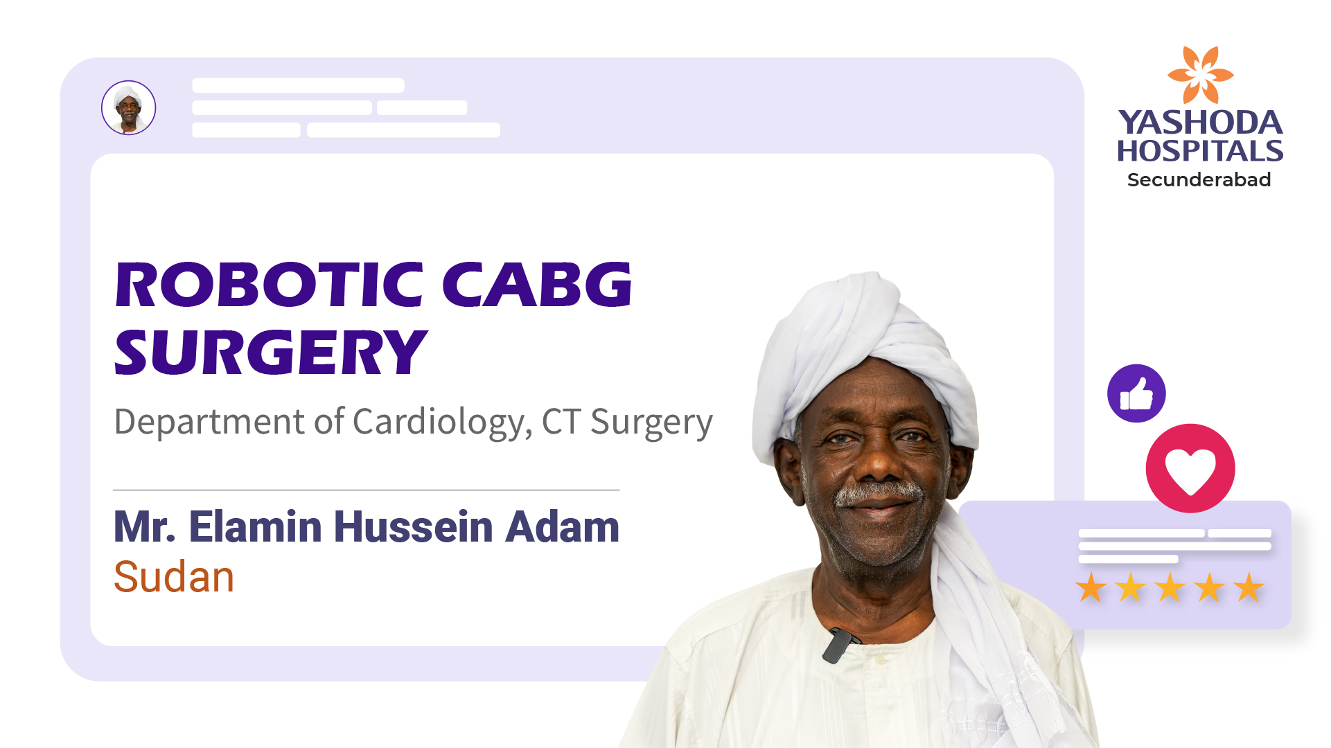
Coronary artery disease (CAD) is a serious condition where coronary arteries become narrowed or blocked, primarily due to atherosclerosis. This…
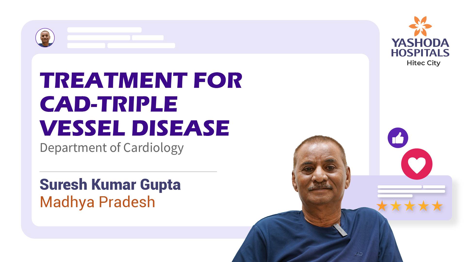
Coronary artery disease (CAD) and triple vessel disease (TVD) are heart conditions caused by atherosclerosis, a buildup of plaque in…
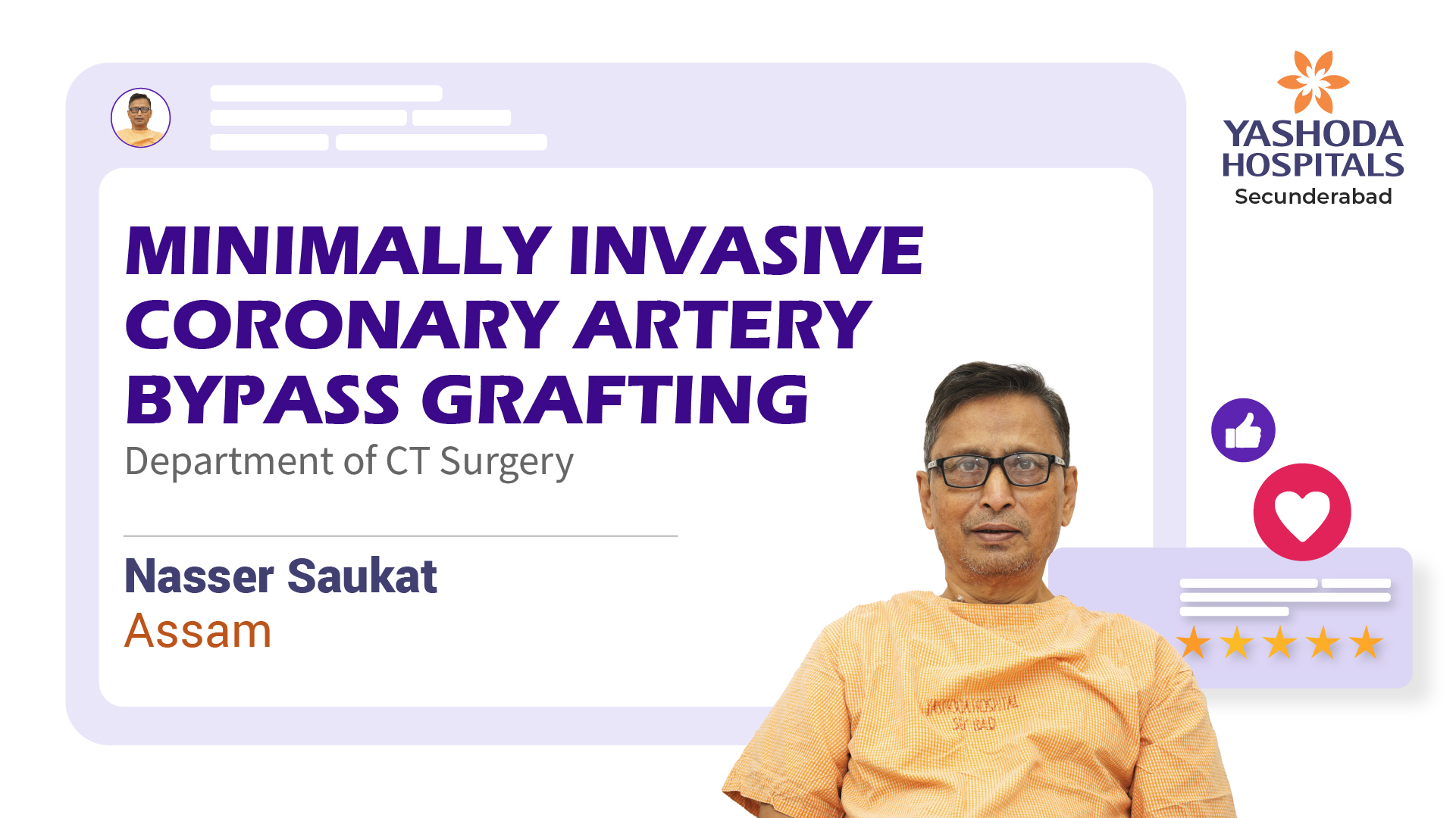
Left Main Coronary Artery Disease (LMCAD) and Triple Vessel Disease (TVD) are serious conditions characterized by extensive coronary artery disease.…
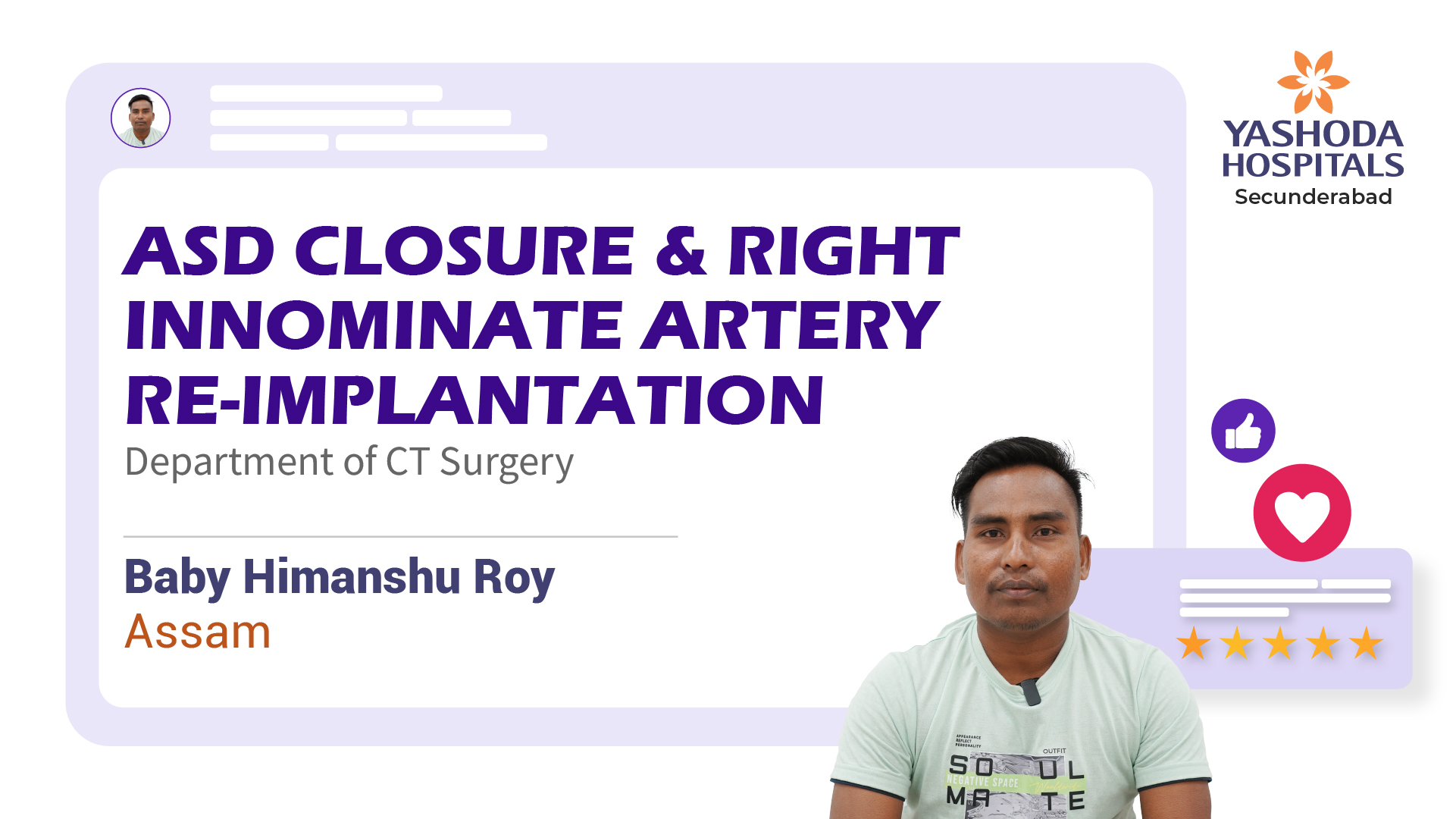
Atrial Septal Defect (ASD), also known as a hole in the heart, is a congenital heart defect where an abnormal…
Unstable angina is a condition where the heart muscle doesn't receive enough blood flow, often a warning sign of an…
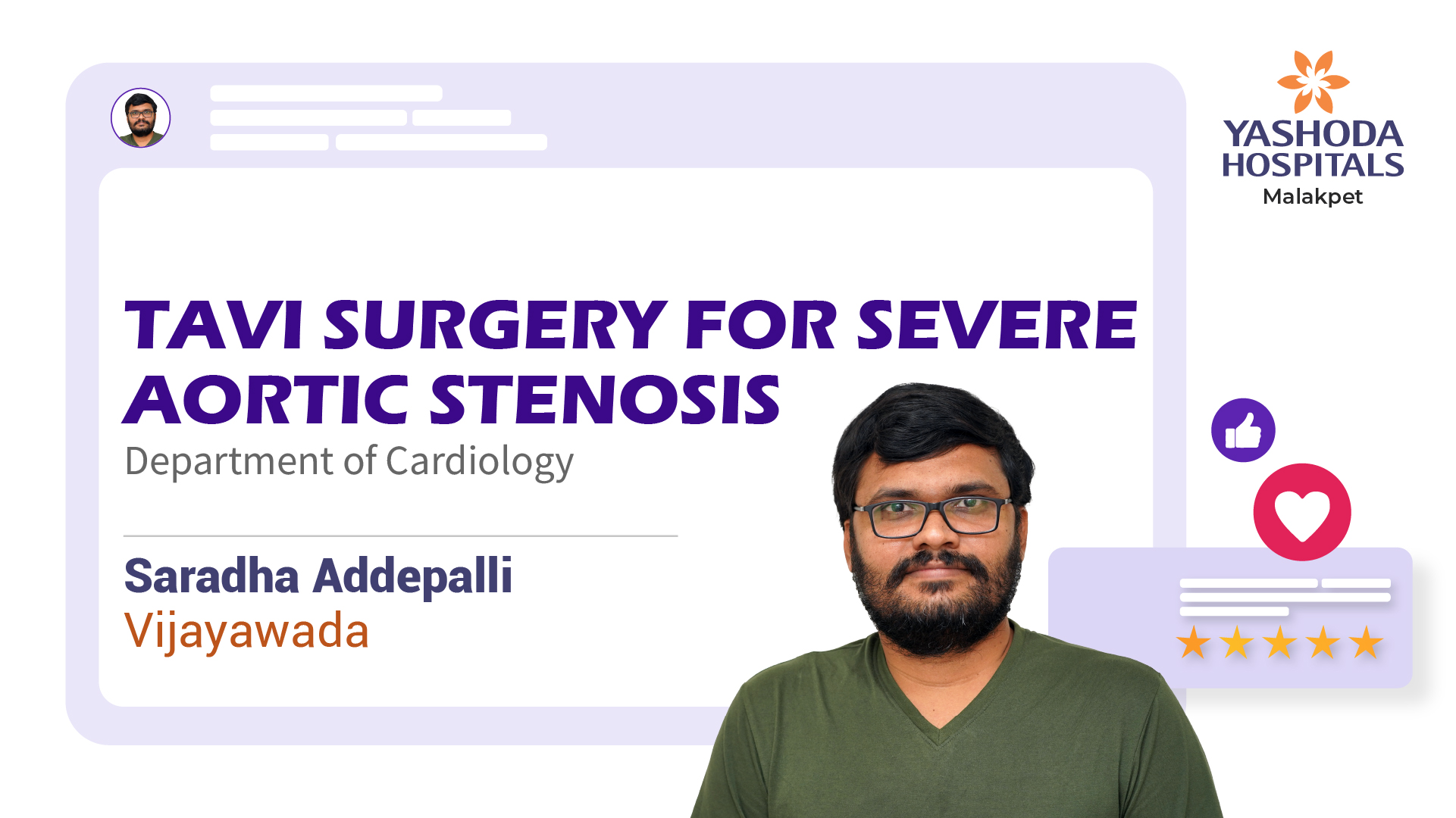
Severe aortic stenosis is a heart condition characterized by a significant narrowing of the aortic valve, obstructing blood flow from…
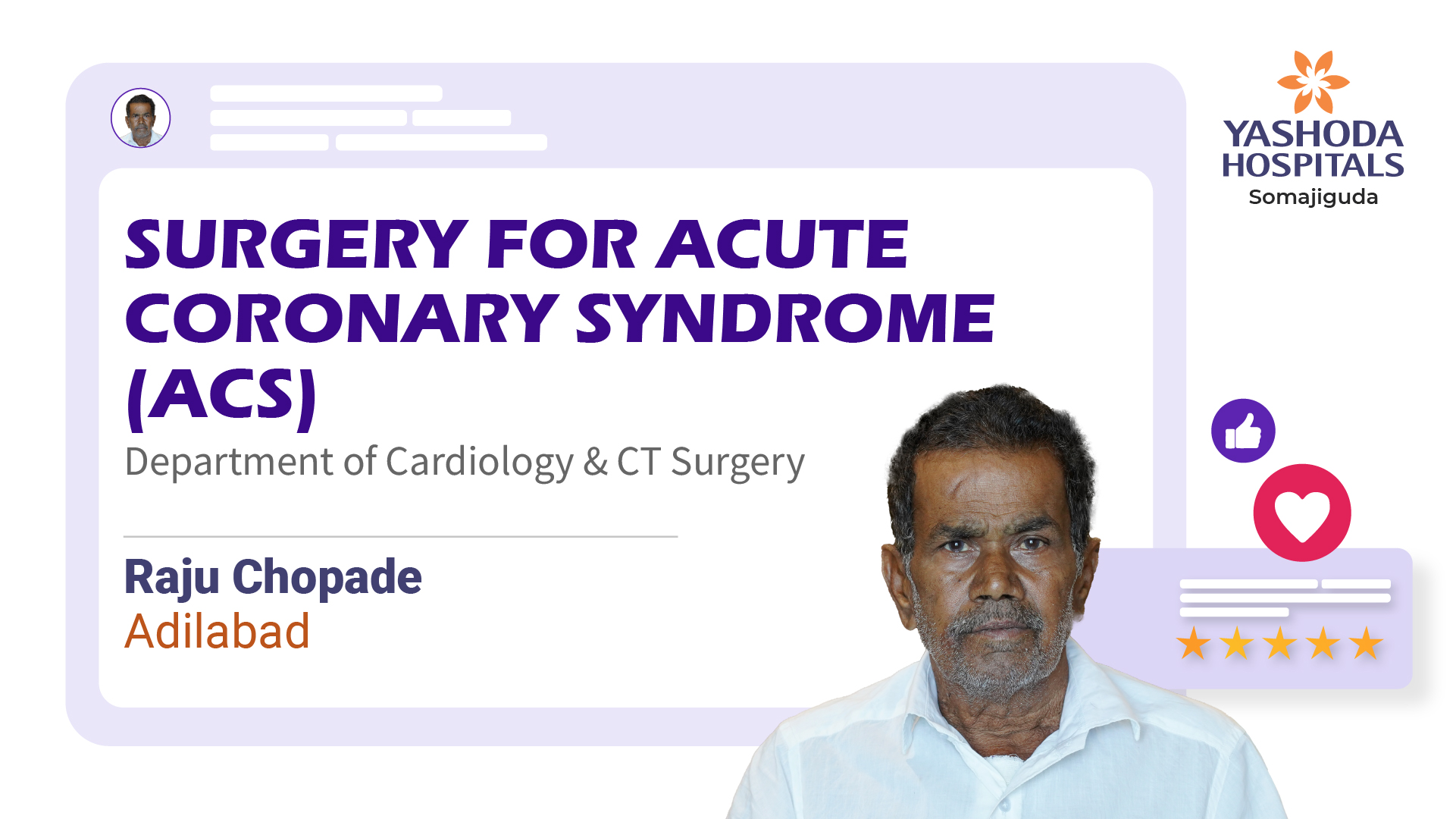
Acute Coronary Syndrome (ACS) is a condition resulting from a sudden reduction in blood flow to the heart muscle, often…
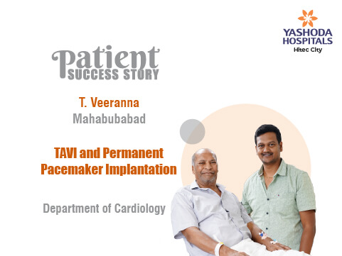
Transcatheter Aortic Valve Implantation (TAVI) is a minimally invasive procedure used to replace a damaged aortic valve in the heart.…
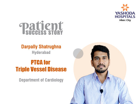
Triple Vessel Disease is a type of coronary artery disease in which the major blood vessels that supply blood to…
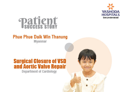
Phue Phue Daik Win Thanung from Myanmar successfully underwent Surgical Closure of VSD and Aortic Valve Repair at Yashoda Hospitals,…
Health Blogs for Cardiology
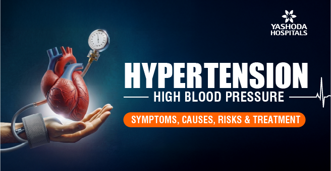
Hypertension (High Blood Pressure): Symptoms, Causes, Risks & Treatment
Apr 08, 2026 05:56Learn about hypertension — its symptoms, causes, risk factors, and treatment options. Discover how to manage high blood pressure and reduce health risks effectively.
గుండె వైఫల్యం : కారణాలు, ప్రమాద కారకాలు, లక్షణాలు, చికిత్స, జాగ్రత్తలు
Sep 25, 2025 10:50గుండె మన శరీరంలో అత్యంత ముఖ్యమైన అవయవం. మన శరీరంలో ఉన్న అన్ని అవయవాలకు రక్తాన్ని సరఫరా చేస్తుంది. ఈ పని చేయడానికి గుండె నిరంతరం కొట్టుకుంటూనే ఉంటుంది. అయితే మనం చేసే పనులను బట్టి కొన్నిసార్లు గుండె కొట్టుకునే వేగం పెరగవచ్చు.
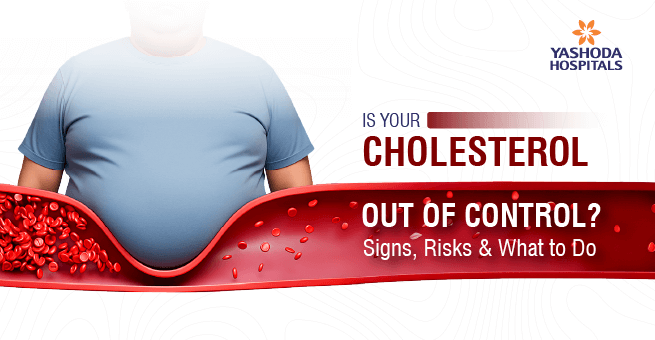
Is Your Cholesterol Out of Control? Signs, Risks & What to Do
Jul 10, 2025 12:05Cholesterol is a natural substance & even necessary. It is often considered a sign of something dangerous, but good cholesterol levels can play a vital role in the body, from helping in the digestion of fatty foods to synthesizing hormones and vitamins, such as vitamin D, and supporting cell membrane integrity.
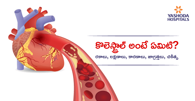
కొలెస్ట్రాల్ అంటే ఏమిటి? రకాలు, లక్షణాలు, కారణాలు, జాగ్రత్తలు, చికిత్స
May 22, 2025 09:06మన శరీరంలో ఉండే రక్తం అనేక రకాలైన కణాలను కలిగి ఉండి నిరంతరం శరీరమంతా ప్రవహిస్తూ ఉంటుంది.
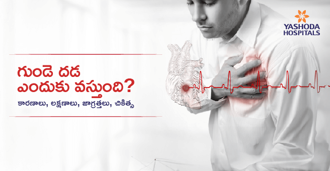
గుండె దడ అంటే ఏమిటి? కారణాలు, లక్షణాలు, జాగ్రత్తలు, చికిత్స
May 08, 2025 06:27ఈ మధ్య కాలంలో గుండెకు సంబంధించిన సమస్యలు ఎక్కువ అవుతున్నాయి, వయసుతో సంబంధం లేకుండా అన్ని వయసుల వారి మీద ఈ ప్రభావం చూపిస్తున్నాయి. కోవిడ్-19 తర్వాత మన ఆరోగ్య పరిస్థితుల్లో చాలా మార్పులు కనిపిస్తున్నాయి.
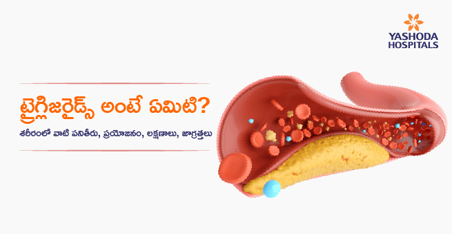
ట్రైగ్లిజరైడ్స్ అంటే ఏమిటి? శరీరంలో వాటి ప్రయోజనం, పనితీరు, లక్షణాలు, జాగ్రత్తలు
Apr 30, 2025 06:55ట్రైగ్లిజరైడ్స్ అంటే రక్తంలో ఉండే ఒక విధమైన కొవ్వు పదార్ధాలు, ఇవి రక్తంతో పాటుగా రక్తనాళాల్లో ప్రవహిస్తూ ఉంటాయి. శరీరానికి శక్తి అవసరమైనప్పుడు తగినంత ఆహారం తీసుకోవడం ఆలస్యమైతే ఆ సమయంలో ట్రైగ్లిజరైడ్స్ ను కరిగించి శరీరం ఉపయోగించుకుంటుంది.
Angioplasty Explained: Here’s What You Should Know
Dec 10, 2024 19:00Angioplasty, also called balloon angioplasty, is a procedure designed to allow the free flow of blood through the arteries. This minimally invasive approach used by cardiologists targets narrow areas due to plaque formation or total blockage of some arteries.

కరోనరీ యాంజియోప్లాస్టీ: రకాలు, ప్రయోజనాలు & సర్జరీ తరువాత తీసుకోవాల్సిన జాగ్రత్తలు
Nov 20, 2024 12:54ఇటీవల కాలంలో గుండె జబ్బుల బారిన పడుతున్న వారి సంఖ్య బాగా పెరుగుతోంది. అయితే గుండె సమస్యలకు ఎప్పటికప్పుడు అధునాతన చికిత్స పద్దతులు అందుబాటులోకి వచ్చాయి. అందులో యాంజియోప్లాస్టీ ఒకటి. గుండెకు రక్తాన్ని సరఫరా చేసే కరోనరీ ధమనుల్లో ఏధైనా అవరోధం ఏర్పడినప్పుడు రక్త ప్రహావానికి అంటంకం కలుగుతుంది. బ్లాక్స్ అని మనం పిలవబడే ఈ అడ్డంకులు లేత పసుపు రంగులో ఉండే ఒక జిగురైన పదార్థం (చెడు కొవ్వు) వల్ల ఏర్పడతాయి.
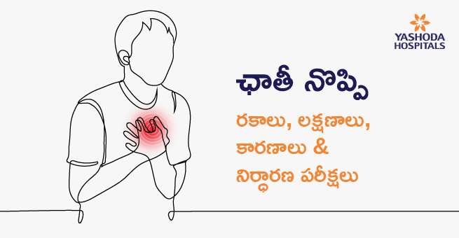
ఛాతీ నొప్పి: రకాలు, లక్షణాలు, కారణాలు & నిర్ధారణ పరీక్షలు
Nov 14, 2024 16:00మనలో ఎంతో మందికి ఎదురయ్యే సాధారణ సమస్యలలో ఛాతీ నొప్పి కూడా ఒకటి. క్రమరహిత జీవనశైలి, చెడు ఆహార అలవాట్ల కారణంగా ప్రస్తుతం చాలా మంది ఛాతీ నొప్పి సమస్యతో బాధపడుతున్నారు. అయితే మనలో ఎంతోమంది ఏదో ఒక సమయంలో ఈ సమస్య బారిన పడే ఉంటారు.
Cardiopulmonary Bypass Surgery: A Life-Saving Procedure for Heart Patients
Sep 30, 2024 16:27Cardiopulmonary bypass (CPB) refers to a procedure that takes place during open heart surgery to temporarily replace the functioning of the heart and lungs. It is a requirement for many sophisticated heart procedures, enabling surgeons to carry out intricate tasks without fear of bleeding or injuring the chest and brain.
Doctor Talk
Health Talk
FAQ’s
What is cardiology?
Cardiology is a branch of medicine that focuses on diagnosing, treating, and preventing heart and blood vessel diseases. Cardiologists handle issues like heart attacks, arrhythmias, and hypertension, among others.
Why should we choose Yashoda Hospitals for Cardiology?
Yashoda Hospitals, recognised as one of the best heart hospitals, offers cutting-edge cardiac care with experienced specialists and advanced treatments, from angioplasty to heart transplants. Our state-of-the-art facilities ensure high-quality care and excellent outcomes.
What are the four silent signs of a heart attack?
Silent signs of a heart attack include unexplained fatigue, discomfort in the back or jaw, shortness of breath, and nausea or lightheadedness. These symptoms can be subtle but need prompt attention.
What should people do to keep their hearts healthy?
To keep your heart healthy, eat a well-balanced diet, exercise regularly, quit smoking, manage stress, and monitor health metrics such as blood pressure and cholesterol.
Where can I find the best heart hospital near me?
Yashoda Hospitals are well-known for their specialized cardiology and cardiothoracic vascular surgery (CTVS) treatments, especially for complex coronary artery diseases, structural heart disorders, and critical cardiac management. You can find one of the most highly-rated heart hospitals near you at the Somajiguda, Hitec City, Malakpet, or Secunderabad branches. They offer a full spectrum of advanced treatment modalities, including interventional cardiology, electrophysiology, and 24/7 emergency cardiac care. Furthermore, patient feedback consistently highlights the department’s ability to provide complex diagnoses, surgical benefits, and essential long-term lifestyle and follow-up plans.
Is heart attack and heart failure the same?
No. A heart attack is a sudden blockage of blood flow to the heart, while heart failure is the heart’s inability to pump blood effectively. These two conditions require different treatment approaches.
What are the treatments available in the Cardiology department?
Treatments in the cardiology department include angioplasty, peripheral vascular interventions, pediatric cardiac care, electrophysiology, pacemaker implants, TAVI, and heart transplantation, among others.
What can I expect during a cardiology consultation?
During a cardiology consultation, you can expect a review of your medical history, a physical exam, possible diagnostic tests, and a discussion of the results with the appropriate treatment plan.
What are the common diagnostic tests used in cardiology?
Common diagnostic tests in cardiology include ECG, echocardiogram, stress tests, cardiac catheterization, and Holter monitoring, among others.
What is the difference between a cardiologist and a cardiac surgeon?
A cardiologist diagnoses and treats heart conditions using non-invasive or minimally invasive procedures, whereas a cardiac surgeon performs complex heart surgeries such as bypasses and transplants.
What is coronary artery disease?
Coronary artery disease (CAD) occurs when your coronary arteries, which supply oxygen-rich blood to your heart, become narrowed or blocked due to cholesterol buildup. Often called a silent killer, CAD may not show symptoms until a blood clot forms, blocking blood flow and leading to a heart attack. It’s possible to have CAD for years without knowing, as symptoms typically emerge only during a heart attack. CAD is also known as coronary heart disease (CHD) or ischemic heart disease.
How to treat coronary artery disease?
Treatment of Coronary artery disease typically involves lifestyle changes, risk management, and medications, with some cases requiring surgery. Following your treatment plan is crucial to reducing the risk of serious complications.
How to avoid heart attacks?
Maintaining a healthy lifestyle can help regulate your blood pressure, cholesterol, and blood sugar levels, reducing your risk of heart disease and heart attack.
Does low BP cause a heart attack?
Some individuals may experience a sudden drop in blood pressure or have consistently low blood pressure, which could be connected to an underlying health issue. This can be dangerous, as it may indicate that your heart, brain, or other vital organs are not receiving enough blood flow, putting you at risk for a heart attack or stroke.
What is the difference between a heart attack and cardiac arrest?
A heart attack occurs when a coronary artery is blocked, depriving the heart muscle of blood and oxygen, leading to tissue death if untreated. A cardiac arrest happens when the heart stops pumping blood, causing a person to stop breathing.
What is a silent heart attack?
A silent heart attack occurs without the typical symptoms like chest pain or shortness of breath. Instead, people might mistake it for heartburn, the flu, or a strained muscle. Despite the lack of obvious signs, it still involves a blockage of blood flow to the heart and can cause damage to the heart muscle.
What are the 4 stages of congestive heart failure?
Heart failure progresses through four stages: A, B, C, and D. Stage A involves individuals at risk for heart failure without symptoms or structural issues. Stage B, or pre-heart failure, includes those with structural heart disease or risk factors but no symptoms. Stage C marks symptomatic heart failure with current or past symptoms, while Stage D is advanced heart failure, where symptoms severely impact daily life or require frequent hospitalizations.
Can heart failure be cured?
Failure of Heart can’t be cured, but you can improve your quality of life through lifestyle changes, medications, and sometimes surgery.
What causes cardiovascular disease?
Risk factors for cardiovascular disease (CVD) include behaviors like tobacco use, an unhealthy diet, excessive alcohol consumption, and lack of physical activity.
Is hypertension a cardiovascular disease?
Hypertension isn’t heart disease, but it is a significant risk factor for developing it. That’s why it’s important not to ignore hypertension.
How to reduce heart blockage?
Once plaque builds up in an artery, unclogging it isn’t simple. However, a healthy diet, regular exercise, and avoiding smoking can improve cardiovascular health and prevent further blockages. In some cases, medication or surgery may be required.
How is heart disease diagnosed?
Various tests are used to diagnose heart disease, including blood tests and chest X-rays. Additional diagnostic tools include ECG, echocardiogram, cardiac catheterization, CT scan, MRI scan, and exercise or stress tests.
How to cure heart disease?
Treatment for heart disease varies based on its cause and the extent of heart damage. While adopting a healthy lifestyle is crucial, medications may be necessary to manage symptoms and prevent complications if lifestyle changes aren’t enough. In some cases, a procedure or surgery may be required, depending on the type of heart disease and the degree of heart damage.
How to identify heart problems?
Heart disease encompasses various conditions that affect the heart, including blood vessel diseases like coronary artery disease, irregular heartbeats (arrhythmias), congenital heart defects, heart muscle diseases, and heart valve disease. Many of these conditions can be prevented or managed through healthy lifestyle choices.
What is the best treatment for cardiovascular disease?
Cardiovascular disease treatment varies based on the cause and extent of heart damage. Adopting healthy lifestyle habits, such as following a low-fat, low-salt diet, exercising regularly, getting sufficient sleep, and avoiding smoking, plays a crucial role in managing the condition.
Which hospital is best for Cardiovascular Treatment in Hyderabad?
For over three decades, Yashoda Heart Institute has been a leader in cardiac care in Hyderabad, consistently pushing the boundaries of medical technology and treatment. As one of India’s premier cardiology hospitals, it has achieved several significant milestones, including being the first in the region to perform a simultaneous heart and lung transplant and successfully completing over 20,000 minimally invasive cardiac procedures each year.

 Appointment
 Second Opinion
 Call
Call More
More


