Endobronchial Ultrasound (Ebus)

Interventional pulmonology has revolutionized the field of pulmonary medicine by providing the most advanced minimally invasive procedures to diagnose and treat both malignant and non-malignant disorders of the lungs and airways.
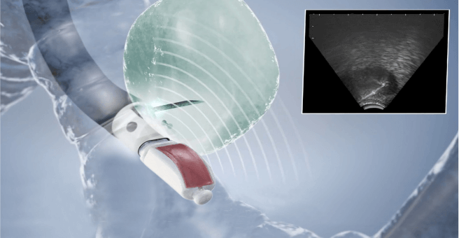
The role of EBUS is progressively expanding to include the evaluation of peribronchial lesions, pulmonary nodules, and other mediastinal abnormalities. Endobronchial ultrasound (EBUS) combines bronchoscopy with ultrasound to enhance the definition of mediastinal structures and aid in the visualization of peribronchial anatomy, reducing biopsy sampling errors due to superior site selection. For transbronchial sampling, EBUS is a minimally invasive but highly effective procedure that provides rapid on-site results and has a very safe profile. It has been shown to be significantly cost-effective compared with prior gold standard techniques.
How is it different?
- An endobronchial ultrasound is much less invasive.
- The physician can perform needle aspiration on lymph nodes using a bronchoscope inserted through the mouth.
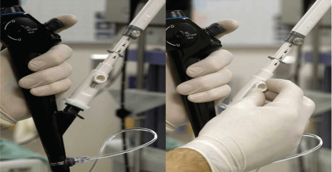
- A special endoscope fitted with an ultrasound processor and a fine-gauge aspiration needle is guided through the patient’s trachea.
- No incisions are necessary.
What are the benefits of EBUS?
- It provides real-time imaging.
- The accuracy and speed of the EBUS procedure allow for rapid on-site pathological evaluation.
- Opportunity for immediate surgery if necessary.
- No cutting or incisions are required.
- EBUS is performed under moderate sedation or general anesthesia.
- Patients may go home the same day if no further surgery is required.
Yashoda Hospitals is one of the largest quaternary centers for Pulmonology & Critical Care in India, with three decades of healthcare presence. We specialize in treating high volumes of pulmonary critical care patients, particularly through minimally invasive procedures, ensuring the best outcomes for our patients.


 Appointment
 WhatsApp
 Call
Call More
More

