Paratesticular Tumor (Leiomyosarcoma)
Background
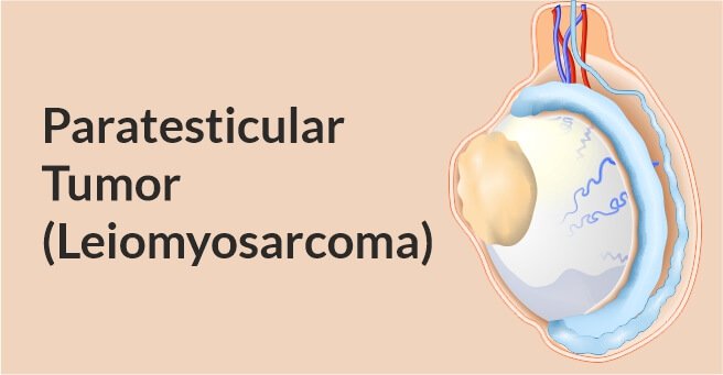
A 61 year old male, non-diabetic and non-hypertensive presented with long standing painful right sided scrotal swelling which gradually increased in size from last 4 months.
Diagnosis & Management
He had undergone ultrasound of scrotum which showed hypoechoic lesion adjacent to right testis. Hypoechoic lesion in right inguinal canal and right hydrocole. He underwent a transcrotal right epididymo-orchidectomy from where a nodular white mass measuring 3.5X2.5 cms was excised.
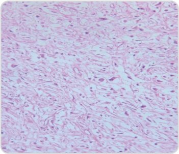
Histopathological examination showed small to large pleomorphic hypoechoic nuclei with moderate to abundant eosinophilic cytoplasm, atypical mitosis noted. Bizzare giant tumor cells were also seen. Benign spindle cells seen-suggestive of Pleomorphic Sarcoma.
HC-showed
- Desmin – Positive
- Smooth Muscle Actin – Positive
- Myogenin – Faint Positive
- Calretinin – Negative IHC MARKERS – favours Leiomyosarcoma
Patient was referred to us for further management.
Blocks were reviewed in our hospital which showed abundant eosinophilic cytoplasm, irregular nuclear borders, vesicular chromatin and prominent nucleoli. Lymphovascular emboli was not seen – suggestive of Pleomorphic Sarcoma.
- Mygenin – Negative
- SMA and Desmin – Positive -favours Leiomyosarcoma
Tumor was infiltrating adjacent fat and margins could not be commented upon
Abundant eosinophilic cytoplasm,irregular nuclear borders vesicular chromatin and prominent nucleoli.
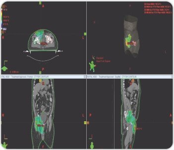
Radiotherapy planning target volume covering post operative bed and regional lymphatics
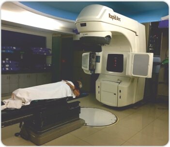
The patient was advised for adjuvant Radiotherapy in view of R1 resection. Patient is on radiotherapy and is on follow up. Initial plan was upto 46Gy covering tumor bed and regional lymphatics, then boost volume covering tumor bed upto 60Gy in view of tumor margins. (Treatment duration: 6 weeks).
About Author –
Dr. Mohanty
MD (Radiation Oncology),
PDCR Consultant Radiation and Clinical Oncologist,
Yashoda Hospitals, Secunderabad



 Appointment
 WhatsApp
 Call
Call More
More

