Robotic lobectomy Surgery for lung adenocarcinoma in a 76-year-old grandmother
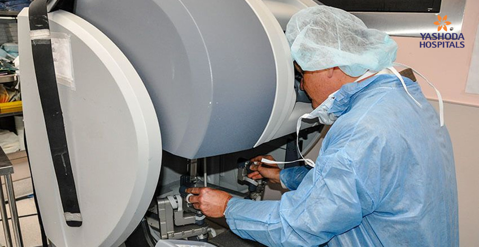
I am thankful to the patient and her family who have consented for me to share this story.
Lung cancer continues to be a life-threatening disease as the survival statistics are not very promising. Surgery is the major treatment for patients with lung cancer (or tumor) to improve their chance at life.
We received an elderly female patient aged 76 years in the thoracic division. Other than a persistent cough, the patient showed no other symptoms. After a series of physical examination and imaging tests, the doctors identified adenocarcinoma in the right, lower lobe of the lungs. The patient required immediate surgery to curb the tumor from progressing beyond stage 2, and spread to other lung tissues or lobe or infiltrate into the lymph nodes and rest of the body.
Ever wonder how lungs look like?
Lungs are a pair of cone-shaped, spongy organs inside the chest. Of the pair of lungs, the right lung is larger and heavier. The lungs have sections called lobes – the right lung has three lobes and the left lung has two lobes. These lobes contain small pockets called alveoli which receive air from the nose through the bronchus and bronchioles. Alveoli are arranged in a branched array very similar to that of the tree branches. Alveoli are the spaces where the air exchange (oxygen/carbon dioxide) takes place and thus, is the vital component of the respiratory system.
Our patient suffered from adenocarcinoma of lungs in the right, lower (inferior) lobe. Adenocarcinoma is the cancer that begins in the glands. Adenocarcinoma of lung, also known as pulmonary adenocarcinoma accounts for 40% of all type of lung cancers.
Complete removal of tumor while protecting the surrounding healthy tissues is important for survival and faster recovery. Also, the lung capacity and respiratory function play a major role in improving the respiratory and overall quality of life.
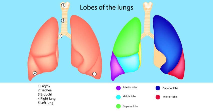
Lungs and its Lobes
Why robotic lobectomy surgery was chosen over video-assisted thoracoscopic surgery?
The major challenge in removing the tumor was its location. Operating the chest area close to the heart is an effort undertaken when benefits outweigh risks. The process of removing tumor from the lungs involves surgical navigation through the vital organs and their life supplies. Usually, the tumor is removed by an open, Video-Assisted Thoracoscopic (VATS) surgery. In this case, however, the risk of morbidity associated with open surgery was too high for the 76-year-old elderly patient, and thus open surgery was not a safe option.
The team was gearing up to deal with vital structures including, heart, pulmonary vessels (vessels connecting blood flow from the heart to lungs for oxygenation) and major nerves in the chest area. For the surgery to be successful we had to achieve some important milestones. Firstly, we had to remove the tumor safely and completely for the patient to be cancer-free. Secondly, major blood vessels and nerves had to be restored in the operated area to maintain the respiratory functions intact.
Navigating through the intricate structures within the lobes to precisely cut-off the tumorous margins and preserve vital structures required greater sophistication and expertise. Even a seasoned cardiothoracic surgeon cannot rule out the possibility of unintentional damage or untoward complications. We had to be extra sure to keep the patient safe and tumor-free.
How robotic assistance enhanced our confidence?
Bleeding is a part and parcel of any surgery, and surgical removal of tumor is no exception. However, in this cardiothoracic surgery for tumor removal, even a small bleed could result in serious complications. We needed technological assistance to remove the tumor without affecting the healthy neighboring structures. Robotic system and visual guidance offered the level of sophistication and precision the surgery needed.
- The robotic system with5mm to 8mm tools offered slow, tremor-free, 1800 rotational movements along the intricate structures in the operation area. The action of robotic arms and the connected tools were easily controlled by the surgeon. As a result, the robotic assistance ensured that all the surgical actions were under control, precise and safe.
- Magnified visual guidance from the built-in camera provided real-time visuals of the pumping heart, nerves, vessels andthe tumor in the lungs. The 10X magnification offered a clear view and navigation guidance through the deep, hidden anatomies.
The high precision robotic arms and real-time visuals meant easier resection of the tumor, safe stapling of the bronchus and blood vessels (to remove the diseased area), and lesser damage to the vital structures in the process. Little or no human handling of the operation area also meant that the patient had reduced risk of infections.
To facilitate an intra-operative biopsy, a medical grade dye, Indocyanine green was injected into the veins of the patient. The real-time visuals showed the dye-laced nodal sites which now appeared with a distinct, green glow. Guided by these real time visuals, we safely performed intra-operative biopsy of the nodes. The biopsy reports confirmed that the tumor was confined and had not spread to the nodes.
Surgeries leave the patient with scars from the incisions (or cuts). Open surgeries especially, are associated with major scars resulting in increased risk of infection and extended recovery time. In robotic lobectomy, surgical incisions as small as 8 to 10 mm were performed; these are very small when compared to approximately 10 cm incisions made during open surgeries. As a result, the patient had lesser blood loss and most importantly retained her cardiac and respiratory functions intact. The patient was in the recovery for just four days and was discharged soon after.
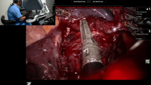
Robotic Surgery stapling
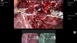
Intraoperative biopsy
How is the patient doing now?
Robotic lobectomy is a major surgery, yet the patient did not suffer any post-operative complications. We are happy that the patient, especially being elderly, had a faster recovery from tumor and surgery. Robot-assisted surgery substantially reduced the respiratory morbidity from the tumor she had. She is now tumor-free and enjoying the new lease of life.
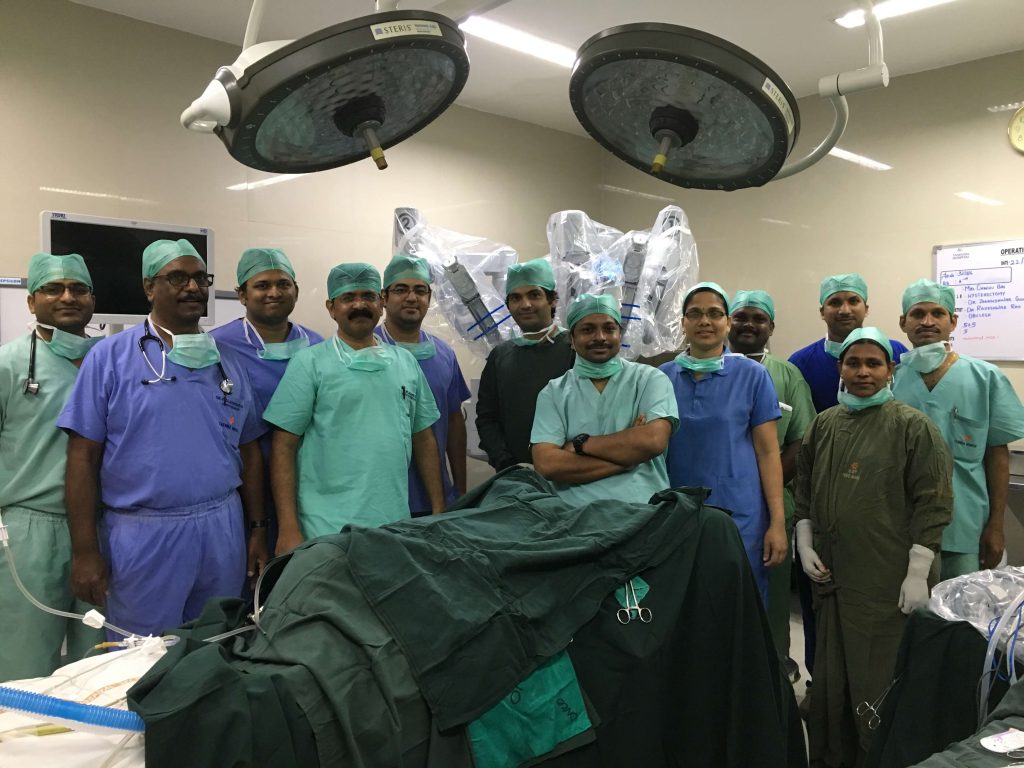
Robotic Surgery Team
For every doctor, it is a hearty satisfaction when the patient has healed to such an extent. While the surgery can guarantee successful remission, the advances in the robotic science and surgical technology continue to redefine “What is possible?”! We have come a long way from the days when cancer was considered to be a final death row. There is growing hope for tumor patients with further advances in the days to come. There is still hope for more and more tumor survivors to create such stories.
About Author:
HOD, Surgical Oncology | Clinical Director Robotic Surgery, Yashoda Hospitals, Hyderabad
His expertise includes robotic surgeries, video-assisted thoracoscopic surgeries, and laparoscopic surgeries. Esophagectomy with nodal dissection, lobectomy, and low anterior resection are among the several surgeries he performs on regular basis.



 Appointment
 WhatsApp
 Call
Call More
More

